Buy Needles And Syringes With No Prescription
M4B Store Banner
intex
Riptropin Store banner
Generation X Bodybuilding Forum
Buy Needles And Syringes With No Prescription
Mysupps Store Banner
IP Gear Store Banner
PM-Ace-Labs
Ganabol Store Banner
Spend $100 and get bonus needles free at sterile syringes
Professional Muscle Store open now
sunrise2
PHARMAHGH1
kinglab
ganabol2
Professional Muscle Store open now
over 5000 supplements on sale at professional muscle store
azteca
granabolic1
napsgear-210x65
esquel
over 5000 supplements on sale at professional muscle store
ashp210
UGFREAK-banner-PM
1-SWEDISH-PEPTIDE-CO
YMSApril21065
over 5000 supplements on sale at professional muscle store
advertise1
tjk
advertise1
advertise1
over 5000 supplements on sale at professional muscle store

Dat's - CJC-1295 & GHRP-6 (Basic Guides)

likkayouth said:
Do you remember how old that someone was?

Dude, a 25 year old girl to you is an "older woman". :p

I know Big A has a rule against giving direct advice but I'm going to do it anyway.

Go hook up with a "Mrs. Robinson" for the next few semesters. Trust me your whole perspective will change...
 
Dat I remember you said that fat has a bigger negative impact on ghrp6 than carbs, so with carbs you wait about 10min aftewards to take your shot, but I remember you saying you take your bedtime shot about 30min after a protein/fat meal/shake. Now, why take the Ghrp6 30min after that meal? Wouldnt it be better to take it 30min before the meal? B/c only 30min after the fat is pretty active right?
 
Dat I remember you said that fat has a bigger negative impact on ghrp6 than carbs, so with carbs you wait about 10min aftewards to take your shot, but I remember you saying you take your bedtime shot about 30min after a protein/fat meal/shake. Now, why take the Ghrp6 30min after that meal? Wouldnt it be better to take it 30min before the meal? B/c only 30min after the fat is pretty active right?

Nope you remembered incorrectly. I take my GHRH+GHRP-6 probably 2.5 hours after my last meal but 20 to 30 minutes before my next meal.

So specifically to your questions:

Q: why take the Ghrp6 30min after that meal?
A: Don't do that.

Q: Wouldnt it be better to take it 30min before the meal?
A: Yes. That is what I do and I believe I always recommend.

Q: B/c only 30min after the fat is pretty active right?
A: GHRH+GHRP-6 act within the first 5 minutes to begin GH release. This continures and peaks between 15 and 30 minutes, After the GHRH+GHRP-6 has "done its thing to create GH" EAT.
 
Anti-aging - GH, GHRH & Ghrelin-mimetics (i.e. GHS)

ANTI-AGING

What follow is a back-bone against which we may come to understand aging and hormonal replacement & associated methodologies that lead to restoration of youthful bodily function in conformance with physiology. It is an understanding that recognizes and attempts to achieve a balanced physiological conformance rather then the imposition of solutions. The body is far more intelligent then the clever solutions we attempt to impose on it.

This will be a long read and will be broken into several postings and many of you may not have an interest. But by understanding and utilizing the concepts already discussed in this thread in specific regard to Growth Hormone Releasing Hormone and Growth Hormone Releasing Peptides we have now taken a significant step toward properly remeding one aspect of the aging "problem".

I fully expect that in a matter of time that we will have properly remedied the remainder. We live in very exciting times in terms of discovery. It is up to us to add daily to our knowledge base and attempt to apply that knowledge to reducing the effects of aging.

The following is directly derived from,
Molecular Endocrinology and Physiology of the Aging Central Nervous System, Roy G. Smith, Lorena Betancourt, and Yuxiang Sun, Endocrine Reviews 26(2):203–250 2005

Most of the sections in the paper are excluded. The parts specifically related to Growth Hormone are included as well as my comments and additional cutting edge material.

If after reading what follows you have an interest in a deeper understanding of Age-Dependent Changes in Biological Rhythms, Aging, Memory, and Cognitive Decline, the Hypothalamic-Pituitary Gonadal Axis and Aging, Sexual Behavior and Aging, HPA Axis and Aging and Transcriptional Regulation and Aging, I encourage you to read the full article. **broken link removed**

From the CONCLUSION and SUMMARY

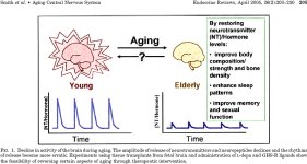
The hormones having the most significant pivotal roles in the Central Nervous System are estradiol, testosterone, cortisol, GH, and IGF-I. In addition to their interplay with neurotransmitters and the complexity of their interactions, they act upstream of the most important regulators of neuronal function. Not surprisingly, hormone replacement has been exploited to improve and maintain quality of life in aging subjects by attenuating the decline in sexual function, memory, learning, mood, quality of sleep, and physical abilities. However, today’s hormone replacement therapies, with the exception of GHS-R agonists for the GH/IGF-I axis, are not ideal because they fail to restore the physiological hormone profile of young adults.

[We have taken a huge step forward in our understanding of hormonal restoration in this thread by understanding that we have now achieved what most thought not possible. That is a youthful restoration of the GH/IGF-1 axis in a manner that conforms to the physiological nature of the body though the use of GHRH and GHRPs.]

Realization of the complex interdependence of the regulatory pathways involved emphasizes the limitations of reductionism. The application of chaos and complexity theories, as introduced briefly in Section II, to biological aging should provide new tools for hypothesis testing and accelerate our understanding of the molecular endocrinology of aging of the CNS.

[Section II reproduced below gives us a framework to view the aging body.]

In the search for a CNS receptor that would restore hormone pulsatility in the elderly to the physiological profile observed in young adults, a group at Merck developed synthetic agonists and then characterized and cloned the orphan GHS-R that regulates the pulse amplitude of GH release (25, 276–278, 280). Following cloning of the GHS-Receptor, an endogenous hormone, ghrelin, was discovered (92). The discovery of the GHS-R established a precedent for the discovery of CNS receptors: they are pivotal regulators of physiological centers affected by aging.

Agonists of the GHS-R reverse physiological aging of the GH/IGF-I axis partially restore thymic function, increase bone density, are neuro- and cardioprotective, and attenuate production of inflammatory cytokines (25, 201, 280, 315, 557–559). Based on this precedent and renewed emphasis on aging research, we are optimistic that methods of endocrine intervention to delay or prevent age-related endocrine and CNS changes that precede detrimental effects on function in the elderly will be forthcoming.

To understand the biological basis of functional aging, it is critically important to combine a systems approach with reductionist methods. Most of the published work we discussed describes correlations rather than causal relationships. Our challenge is to design experimental paradigms with a biological systems approach in mind for incorporation into a neuroendocrinology model of aging. Such an approach will allow identification of pivotal points in aging pathways that lend themselves to intervention.

So lets delve into the paper and look at what we know & where we need to go.

Fig1.jpg

II. Complex Behavior of Neurons in Aging

A. Aging and the dynamics of neuronal behavior

What underlying principle might explain the progressive physiological changes that lead to an "old" phenotype? In general, physiology is governed by complex interactions arising from feedback loops of nonlinear systems; it has been proposed that a reduction in the complexity of physiological or behavioral control systems occurs with age and disease (39–44). A hypothetical advantage for biological systems to exist at the "edge of chaos" is that it allows synchronized neuronal networks to be more resistant to disruption than systems with either periodic or stochastic behavior. Hence, the nonlinear dynamics of ordered chaotic systems facilitates neural systems to adapt according to environment (40, 41). The aging phenotype reflects reduced ability of an organism to adapt to stress and trauma, which is consistent with a transition toward reduced complexity of the underlying regulatory systems.

[It is our ability to adapt and evolve in response to environmental inputs that allow us to survive AND act intelligently. A completely non-chaotic system is one of order. A rock represents an ordered system. A completely chaotic system has no order and thus no predictability. We as humans are systems that are constructed in such a way as to behave in ways that lie between the two extremes of oder and chaos. We are in essence complex.]

Reduced complexity could occur through loss or defect in a component and/or altered nonlinear coupling (feedback) between components of the system (45). A loss of neuronal components and coupling between components of neuronal networks is characteristic of aging. For example, a relative increase in the concentration of glucocorticoids compared with sex steroids, GH, and IGF-I is associated with shrinkage of the hippocampus, loss of neurons, and declining neurogenesis; loss of estradiol production is associated with fewer neuronal connections. The number of dopaminergic neurons also gradually declines during aging, producing deficits in the nigrostriatal dopamine system of rodents, monkeys, and humans (19, 46, 47). Such changes would predictably result in reduced complexity and efficiency of signaling within neural networks and reduced adaptive capability. An example of a decline in adaptive capacity of neurons during aging is the increased vulnerability of the brain to anoxia and ischemia, which in rats is associated with reduced glycolytic capacity of neurons (48). On this basis, we speculate that the onset of functional deficits associated with aging is a consequence of altered behavior of underlying regulatory pathways in the CNS.

[So the aging adult begins to loose some of that complexity of youth becoming less dynamic and responsive to change. The mini-evolutions that take place become less frequent in large part because of the lossed/reduced capacity of our components to communicate with each other.]

B. Hormone pulsatility and aging

One of the most significant age-related events is an alteration in amplitude and pulsatile pattern of hormone release. The frequency of release of a hormone is as important, or more important in some cases, than the amount of hormone released. Target cells respond most effectively to exogenous hormonal stimulation when the frequency of stimulation approaches the endogenous frequency (49). Age-related changes in the endocrine system can appear superficially as apparent increases in complexity (45, 50–54). Veldhuis and colleagues (27, 55–58) made extensive evaluations of age related changes in the dynamics of pulsatile hormone release. They applied mathematical approaches to investigate the synchrony and pulsatility of GH, LH, testosterone, ACTH, cortisol, and insulin release during aging. By calculating the approximate entropy (ApEn) statistic as a measure of orderliness of synchronicity of hormone release, they showed that individual orderliness declined progressively during healthy aging. However, ApEn calculations do not directly distinguish between contributions of stochastic and deterministic behavior toward the observed regularity (45, 53). Therefore,the less ordered rhythmic patterns of hormone release observed during aging could result from a transition of the regulatory neuronal network controlling the ordered frequency of hormone release from adaptive complex behavior to stochastic behavior.

The ApEn calculations in concert with clinical data support the concept that aging is tightly associated with disruption of the time-delayed positive and negative feedback pathways controlling synchrony of hormone release. Therefore, application of nonlinear dynamics and mathematical analyses for analyzing the behavior of neurons that regulate the endocrine system and how this behavior changes as a function of age is important and reinforces our awareness of the limitations of reductionist methods.

[Pulsatility of hormonal release is important because as I have indicated before it is a signaling component, or a method of communication and really nothing more. Therefore the characteristics of generated pulses are very important in communicating how the central nervous system expects cellular structures to behave. The system and how components relate to each other is what seems to be of utmost import, rather than precisely what happening within a component.]

C. Dopaminergic system as an example of age-related change in neuronal dynamics

In aging rats, dopamine production decreases as reflected by reductions in tyrosine hydroxylase mRNA (TH) in the pars compacta of the substantia nigra, and ventral tegmental area (59). The number of cells expressing TH mRNA is the same in young and old rats, but aging is associated with reduced TH gene expression per cell. Reduced production of dopamine during aging increases the susceptibility of neurons toward glutamate neurotoxicity, resulting in seizures and neuronal cell death (60). Dopaminergic neurons in the caudate-putamen, substantia nigra, and nigrostriatal pathway also show increased susceptibility to degeneration induced by 1-methyl-4-phenyl-1,2,3,6-tetrahydropyridine treatment (61). This gradual loss of neurons from the neuronal network will be accompanied by progression toward reduced complexity in neuronal behavior.

Studies of the behavioral dynamics of the dopamine neurons are consistent with age-related progressive changes toward reduced complexity. When the electrophysiological behavioral characteristics of dopaminergic neurons were compared in the brains of young and old animals, two different firing modes (single-spike and bursting) that interweave to produce irregular interspike patterns were identified (62– 65). Mathematical analysis to discriminate nonlinear deterministic from either stochastic or linear oscillations showed that interspike intervals recorded from dopaminergic neurons exhibited a transition toward stochastic behavior during aging (65, 66). Although irregular stochastic behavior could also organize the irregular behavior of neurons, the rapid synchronization and processing of irregular input signals is less readily accommodated (41).

[Changes in the firing pattern of cells between the young and old seems to be a central theme. But what happens at least in regard to somatotroph cells is far more interesting then what is described here. Somatotrophs self organize and create three-dimensional networks that are tightly ordered complex structure which become less so as we age. Proper functioning of the GH cell population as a whole depends on the fine tuning of the number, activity, and positioning of GH cells and this occurs by very rapid inter-cellular communication. See the study posted below.]
 
Last edited:
Anti-aging - GH, GHRH & Ghrelin-mimetics (i.e. GHS)

ADVANCED TOPIC - Pulsation

Heretofore we have discussed the mechanisms involved in effecting growth hormone secretion in terms of a trilogy of hormones acting on the pituitary. We have noted that the pulsatile release of growth hormone is very important in effecting growth. We have intimated but not yet discussed the signaling events that occur once a growth hormone ligand binds to a receptor and how these events effect the transcription of proteins inside the cell nucleus. We have discussed growth hormone, IGF-1 and insulin in terms of circulatory levels but we have not examined the extent to which these respective ligand-bound receptors interact with one another to effect protein transcription and in particular tissue growth.

Eventually we may delve into some of those aspects. But before we do it is important to actually understand pulsation.

Hormonal pulsation is incredibly important. Pulsation conveys hormonal signals which upon ligand receptor-binding transmits information content to regulators of signaling pathways which go on to effect intracellular happenings. For example one signaling pathway, Extracellular-signal-regulated kinase (ERK) is effected by three categories of regulators which act in accordance with the instruction set received from the pulse.

The "temporal regulators" control duration of ERK activation.
The "strength controlling regulators" control magnitude of ERK activation.
The "spatial regulators" control the location of ERK activity, either in cytoplasm or within the cell nucleus effecting transcription.

The end response varies by cell type. For instance if ERK activation is of long duration it triggers proliferation in fibroblast cells but not in PC12 cells. PC12 cells will instead differentiate and only proliferate if the ERK activation is transient.

The specifics aren't important. This example simply demonstrates the importance of pulsation in transmitting information which has diverse effects within various cells.

Conventional View of Pulsation

The conventional view holds that the pituitary possesses growth hormone secreting cells that secrete growth hormone as a response to growth hormone releasing hormone or cease growth hormone release as a response to somatostatin. In this model these cells respond and act individually. They are in many ways "dumb" and possess only the ability to either act or not act.

The addition of Ghrelin mimetics (i.e. GHRPs) adds a third external "modulating" hormone to the model but the conventional view of how somatotrophs (growth hormone secreting cells) behave is unchanged.

But this is not only an overly simplistic model it is incorrect. Yet very few understand the actual mechanism. In fact 99.9% of the scientific community is likely unaware of the research we are about to quote.

The following study provides a new understanding:

Revealing the large-scale network organization of growth hormone-secreting cells, Xavier Bonnefont et al, PNAS, November 15, 2005 vol. 102 no. 46

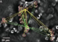
In this study they used three dimensional real-time imaging of the entire pituitary in specially treated mice to look at the positioning and the signaling of every growth hormone secreting cell. This imaging took place across the life span of the mice and revealed for the first time a functional network of somatotrophs that were "wired" to one another across the whole pituitary gland and this network underwent "geometry-driven changes that correlated with the activity of the GH axis".

In other words they discovered a structure of somatotrophs connected by adherens junctions that communicated with one another to coordinate a response to growth hormone releasing hormone (GHRH).

Here is a partial fluorescent imaging of the active network which consisted of numerous intercrossing strands of single somatotrophs with larger clusters of somatotrophs positioned at the intersection of the strands.

**broken link removed**

They discovered a fluid network structure that varied over the lifespan. In both prepuberty and adults the concentration was more centered in the median portion of the pituitary. During puberty the cluster concentration increased in the lateral sections of the pituitary.

They concluded that it is the "geometry of the GH cell system, rather than the global cell density, that is organized in a way that would promote a more coordinated pulsatile release of GH that accelerates growth at puberty." They further suggest that "intact GH clusters facilitate GH responses to secretagogues, because disaggregation of the GH system by enzymatic dispersion of mature male pituitaries led to a GH release response to GHRH 8-fold lower than that seen from large assemblies of GH cells."

2.jpg

We know that GHRH triggers somatotroph intracellular communication mechanisms, but what this study discovered is that GHRH triggered both intracellular and intercellular somatotroph communication mechanisms with different timescales. GHRH spiked calcium (the signaling pathway) which was associated with "a selective increase in cell connectivity between pairs or triplets of neighboring somatotroph cells." However the "overall number of cell pair connections markedly decreased after GHRH stimulation."

"These results suggest that GHRH does not simply affect all cells independently in this region but changes the local pattern of cell–cell communication, resulting in small islets of more highly functionally connected GH cells at some points in the system, interspersed with functionally less connected GH cells. This paradoxical reduction would 'sharpen' the global pulsatile response by minimizing the contribution of less coordinated cell assemblies within the network. Note that the GHRH responses were markedly different in the median zone in comparison with those observed in the lateral zones, indicating that GHRH triggered spatially stereotyped motifs of cell connectivity, which clearly depend on the local geometry of the GH system architecture within different regions of the gland."​

They go on to conclude,

"Moving the focus from single-cell function to whole-organ function is crucial to understanding function at the systems biology level. It is particularly obvious for GH secretion, which clearly requires a high degree of coordination of a large population of individual GH cells to generate a large secretory burst into the blood stream that survives dilution and mixing in the circulation, to deliver a defined pulsatile signal to peripheral tissues....We show that the apparently dispersed secretory elements of the GH system are actually organized to form a structurally and functionally interdependent network and suggest that the proper functioning of the GH cell population depends on the fine tuning of a triplet involving the number, activity, and positioning of GH cells. This triplet functioning allows for fine tuning of communication mechanisms between large numbers of connected GH cells within a network to produce rapid and synchronized hormone pulses in response to physiological needs."​

It is amazing to me that other than a review in TRENDS in Endocrinolgy and Metabolism there has been no published research that has expanded upon this remarkable research. It will come though...such is the snail pace of science.

But what it does for us is it allows us to understand why there is synergy in GH release between GHRP-6 and GHRH in vivo but not in vitro. GHRP-6 I conjecture plays a role in "sharpening" the clustering or somatotrophs which is important as we age.

What seems to happen w/ some disease or ailments is a fragmented less capable networking of secreting cells which leads to weakened or even incorrect down stream messaging. One cell says to other "I didn't quite catch that ...are we suppose to proliferate, differentiate or induce apoptosis?"

There is some indication that this exact system is also responsible for such systems as Luteinizing Hormone pulsation. In light of this study when one happens by a study* such as the one where males with hypothalamic hypogonadism were successfully treated with GnRH 5-20ug/120min subcutaneously via a pump for four months one can't help but to speculate that the network of secreting cells was relearning how to effectively network.

With specific regard to our interests it has become increasingly clear to me that although there are differences in the isoforms of natural GH and the singular synthetic GH form that isn't really what makes natural GH superior. What makes natural GH superior is the pulsation which leads to intracellular signaling and when the pulse is over and there is "silence" a resensitizing of the signaling pathways enabling them to carry the signal anew. This doesn't happen when GH is continuously present. It is pulsation carrying the proper information to the receptor and into the cell cascading into transcription factors that is paramount.

* - Pulsatile GnRH or human chorionic gonadotropin human menopausal gonadotropin as effective treatment for men with hypogonadotropic hypogonadism: a review of 42 cases, Buchter D et al, Eur J Endocrinol 1998; 139: 298-303
 
Last edited:
Anti-aging - GH, GHRH & Ghrelin-mimetics (i.e. GHS)

ANTI-AGING

Continued from, Molecular Endocrinology and Physiology of the Aging Central Nervous System, Roy G. Smith, Lorena Betancourt, and Yuxiang Sun, Endocrine Reviews 26(2):203–250 2005

V. GH Axis

A. Age-associated decline in GH pulse amplitude

Neuroregulation of pulsatile GH release from the anterior pituitary gland secretion has been reviewed (170). The amplitude of the GH pulses is attenuated as we age because of suboptimal signaling from the hypothalamus. This is partially explained by an age-associated reduction in receptor density for the positive regulator of GH release, GHRH (171– 174). Reduced receptor density would require higher levels of GHRH to normalize the pituitary response. Rudman et al. (175) addressed the question of whether GH replacement in the elderly might have functional benefits. They administered GH chronically to elderly men for 6 months and found improvements in body composition and skin thickness that were consistent with reversal of the aging process (175). Unfortunately, adverse side effects such as carpal tunnel syndrome and gynecomastia were relatively common (175). However, the incidence of adverse events is reduced if lower doses of GH are used (176 –178).

It is important to note that GH replacement by bolus injection overrides the episodic physiological profile (179). Biochemical and biological data support the importance of the episodic profile. In the liver, different signal transduction pathways are activated when pulsatile vs. sustained administration of GH are compared (180). Male rats exhibit pronounced high amplitude pulses, whereas in females the profile is flatter and is reflected by distinctly different patterns of GH-regulated gene expression. In males, whole body pubertal growth rate is dependent on GH activation of signal transducer and activator of transcription 5b (STAT5b). In STAT5b knockout mice, males exhibit a female GH phenotype (181).

In humans, GH pulse amplitude and plasma IGF-I levels decline during aging (24, 38). Young adults exhibit a gender difference, in which women have the same pulse frequency as men but with a 2.4-fold increase in burst mass (182). However, these gender differences disappear during aging, and elderly men and women have equally low amplitude GH pulses and reduced IGF-I levels (24, 38). Based on the known properties of GH and IGF-I in vivo, this reduced amplitude, in combination with reduced sex steroid production, likely explains the observed age-dependent change in metabolism, increased fat/lean ratio, decreased muscle strength, reduced exercise tolerance, and increased bone loss. Hence, the functional deficits that result from aging are probably caused by suboptimal signaling from the hypothalamus. An ideal approach for modifying the aging phenotype would be to restore activity of the hypothalamic neurons that control GH pulse amplitude.

A recent study (183) describes the use of DNA microarray chip technology to relate the physiological decline in GH with molecular mechanisms underlying the aging process. Gene expression was compared in the liver of old rats, with or without GH replacement. Of 1000 genes detected in male rat liver, 47 transcripts were affected by aging and about 40% of the differentially expressed genes were normalized by GH treatment. This study is notable and refreshing because the authors evaluated gene expression in the animals after compensating for changes in GH. However, because of age-dependent changes in other hormones, such as sex steroids, and the difficulty of replacing hormones in a way that recaptures the physiology of a young animal, it is impossible to precisely differentiate hormone-dependent from hormone-independent age-related changes. Despite this caveat, studies using DNA microarray analysis of brain tissue from rats that show improvements in memory following GH replacement should be particularly informative.

B. Increase in longevity in GH-deficient rats and mice

It seems reasonable to speculate that restoration of GH release in a way that mimics the physiology of a young adult will provide functional benefits, not necessarily by increasing longevity, but by improving the quality of life. This is based on observations in GH-deficient humans showing that GH increases bone density and improves body composition, cognitive function, cardiac function, and exercise tolerance. However, despite this evidence, the likelihood of achieving beneficial effects by rejuvenating the GH/IGF-I axis physiologically is not universally accepted. One of the reasons is based on laboratory animal studies, where the data suggest that reducing GH, IGF-I, or insulin signaling increases longevity (184 –194). However, these studies do not address quality of life in elderly subjects, which is more important than longevity. Indeed, although caloric restriction has also been shown to improve longevity in a variety of species, a recent, careful study and review of the literature shows that prolonged caloric restriction impairs cognitive function in rats (195).

The evidence for a negative impact of GH and IGF-I on longevity is based largely on studies in mutant mice that are either GH deficient or lack receptors for GH. All of these models exhibit dwarfism, reduced body temperature, and reduced fertility. Certain of these mouse models, such as the Ames dwarf (dw/dw) mouse are GH, IGF-I, prolactin, and thyroid hormone deficient. Although recent studies (188) indicate that the life span of hormone-deficient dwarf mice housed under stress-free laboratory housing conditions is 50% longer than their normal littermates, these observations are unlikely to predict survival in the natural environment. For example, earlier studies indicated that dw/dw mice had markedly reduced life span (45-60 d) and are immunocompromised (196, 197). The reduced longevity was suggested to be a result of more stressful animal housing conditions, typical of 30 yr ago, and poor adaptation to stress (198, 199). Indeed, it has been speculated that the negative effects of prolonged stress, which causes suppression of the immune system by glucocorticoids, are balanced in wild-type animals by the positive effects of GH, IGF-I, prolactin, and thyroid hormone (198, 200).

It is important to recognize the beneficial roles of the GH/IGF-I axis in human physiology. In addition to antagonizing the adverse effects of chronic stress on the immune system, GH and IGF-I may play a similarly protective role in the CNS, thereby potentially improving quality of life. Indeed, Koo et al. (201) reported that restoring GH levels produced beneficial effects on the immune system of old normal mice. They showed that when old mice are treated with an oral GH secretagogue, restoration of GH and IGF-I reversed the age-dependent shrinkage of the thymus and improved T-cell production. The advantage of rejuvenating the GH/ IGF-I axis was illustrated by implanting aggressively growing tumors into the mice. Treatment with a GHS-R agonist reduced the rate of tumor growth, inhibited metastasis, and increased longevity (201).

[NOTE: That deficiency in GH & IGF-1 only leads to longevity in a non-stress environment. The lack of these hormones reduces the ability of the body to effectively manage stress.]

C. GH in the CNS

The characteristics and significance of GH binding in the human brain have been reviewed by Nyberg and Burman (202). In addition to reduced GH release during aging, the concentration of GH receptors in the brain also declines. The highest density of GH binding is in the choroid plexus, with significant binding in the hippocampus, hypothalamus, amygdala, putamen, and thalamus (202). Although GH receptors are widely expressed in the CNS, and anecdotal reports claim that GH improves mood in the elderly, relatively few studies have investigated GH’s functional role in the brain (203). Indirect evidence suggests that GH plays an important role in CNS function. GH-deficient children have an increased incidence of anxiety, depression, and attention deficits, which may contribute to their observed learning disabilities in arithmetic, spelling, and reading compared with age-matched controls (202, 204).GH-deficiency in adults is reported to be associated with reduced energy, unfulfilled personal life, low self-esteem, problems controlling emotional reactions, social isolation, impaired social function, mental fatigue, impaired general and mental health, and deficits in cognitive function (205–211). Markedly reduced GH levels, particularly the integrated nocturnal levels, have also been associated with major depressive illness (212). This may explain the increased incidence of depression and poor sleep quality in the elderly population.

The neuroprotective property of GH was documented in rats. Hypoxic-ischemic damage causes increased GH transport into the brain as manifested by an increase in the number of GH-immunopositive neurons (213). To demonstrate GH binding by immunostaining, the authors went to extraordinary lengths to document specificity. The immunohistochemical evidence showing that GH migrates to the sites of injury following hypoxic-ischemic injury is most persuasive. The authors also demonstrated that icv administration of GH was neuroprotective in the cortex and hippocampus. Thus, the decline in the amplitude of GH secretion during aging likely attenuates GH-mediated neuroprotection.

D. Relationship of GH and IGF-I to age-related cognitive impairment

A comparison was made of the expression of IGF-I mRNA and protein and IGF-I receptor mRNA in the brain of Fisher 344 x BN rats during aging (214). Age-related changes in IGF-I mRNA were not evident in cortical tissue. However, between the ages of 11 and 32 months, IGF-I protein levels were reduced by 36.5% in the cortex. Although IGF-I receptor mRNA concentrations were unchanged, IGF-I receptor binding was reduced by 27% in the cortex and 31% in the hippocampus. When different age groups of rats were compared (6 months vs. 23 months), a modest decrease in IGF-I mRNA was reported in the hippocampus (215). A decrease in IGF-I concentrations and IGF-I binding in the hippocampus is consistent with age-related neurodegeneration.

GH administration increases IGF-I gene transcription in the CNS. Whether a causal relationship between age-associated deficiencies in cognitive function and declining brain IGF-I levels exists is the subject of continuing debate. However, an association is supported by the observation that when IGF-I is administered icv to rats for 28 d, the age-dependent decline in spatial reference and working memory is reversed (216). GH studies in adults with childhood onset GH deficiency showed that GH replacement benefited CNS function (217, 218). Doses of GH that produced supraphysiological levels of IGF-I normalized memory function after 6 months of treatment. Lower doses selected to provide physiological IGF-I concentrations in the blood improved memory function more slowly, but normal function was restored after 12 months of treatment.

Reduced IGF-I levels are characteristic of aging (2, 215, 219–221). Endogenous IGF-I plays a significant role in recovery from insults such as hypoxia-ischemia (222). Neurons die within hours or days following initial injury because of activation of cell death pathways. However, IGF-I with its binding proteins and receptors is induced within damaged areas following brain injury, which suggests that IGF-I plays a neuroprotective role. Administration of IGF-I within a few hours after brain injury confers protection on gray and white matter; by contrast, IGF-I pretreatment is ineffective, probably because of limited intracerebral penetration into the uninjured brain. This important neuroprotective property of IGF-I argues for the maintenance of young adult IGF-I levels during aging.

It has been suggested that IGF-I deficiency could be involved in cognitive deficits seen with aging. In elderly humans (aged 65-86 yr), a correlation between a subject’s performance in the Mini Mental State Examination and plasma IGF-I was reported (223). To investigate this observation in more detail, cognitive functions known to decline during aging were compared with those insensitive to aging. The outcome was consistent with a protective effect of IGF-I on the onset of age-dependent cognitive deficiencies, particularly in speed of information processing (208, 224). Similarly, Dik et al. (225) investigated whether IGF-I was associated with cognitive performance and cognitive decline over a 3-yr period in 1318 subjects, aged 65-88 yr. Although cross sectionally IGF-I was directly related to information processing speed, memory, fluid intelligence, and Mini Mental State Examination score, these statistics were not significant after adjusting for age and other factors. However, analysis in quintiles of IGF-I illustrated a threshold effect of low IGF-I on information processing speed, with lower speed in those subjects in the lowest quintile of IGF-I (<=9.4 nmol/liter) vs. those in the other four quintiles. A low IGF-I threshold was also observed during a 3-yr decline in information processing speed. In conclusion, this study suggested that IGF-I levels below 9.4 nmol/liter are associated with the level and decline of information processing speed.

Serum IGF-I appears to regulate brain amyloid-B (AB) levels (226). During aging, IGF-I levels fall and AB, which is involved in the pathogenesis of AD, accumulates in the brain. Elevations in brain AB levels are found at an early age in mutant mice having low circulating IGF-I. AB burden can be reduced in aging rats by increasing serum IGF-I, and it reflects the ability of IGF-I to induce the clearance of brain AB. This is probably mediated by enhancing the transport of AB carrier proteins such as albumin and transthyretin into the brain. The enhanced uptake is antagonized by TNF-A. IGF-I treatment of mice overexpressing mutant amyloid markedly reduces their brain AB burden; therefore, IGF-I appears to play an important role in modulating brain amyloid levels.

Studies on centenarians showed increased prevalence of dementia in those with lowest serum IGF-I levels (227). Collectively, these results are consistent with a causal link between the age-related decline of GH and IGF-I levels and cognitive deficits, which reinforces the need for continued investigation of IGF-I and CNS function. Ghrelin mimetics have been shown to normalize IGF-I levels in the elderly and to increase IGF binding protein 3; therefore, these compounds may prove beneficial as neuroprotective agents during aging (25, 38). The fact that IGF binding protein 3 levels are also increased suggests that the risk/benefit ratio regarding cancer risk may not be increased by such treatment.

E. Potential mechanisms of GH/IGF-I-mediated neuroprotection

The age-associated decline in GH and IGF-I is likely to cause deficits in functioning of the CNS because both hormones play an important role in vascular maintenance and remodeling. The cerebrovasculature is a source of IGF-I and nerve growth factor (NGF), which are known to play an important role in memory (216, 228–230). During aging, cerebral blood flow decreases and, together with reduced production of sex steroids, correlates with the age-related decline in plasma GH and IGF-I levels. In BN rats, arteriolar density and anastomoses decline markedly between the ages of 7 and 29 months. However, GH treatment produces increases in IGF-I, reverses the age-dependent changes, and increases the number of cortical arterioles (231). These data suggest that preventing the decline in GH and IGF-I during aging would help prevent age-related reductions in vascular density.

The continued viability of adult neurones requires neurotropic factors to support plasticity and provide neuroprotection. A decline in production of such factors probably contributes to the age-related functional deficits that occur in the aging brain. Through its property as a potent stimulator of myelination, IGF-I should protect against the demyelinating effects of increased levels of TNF-A (232). In mouse glial cultures, TNF-A increases apoptosis of oligodendrocytes, whereas IGF-I acts as a neuroprotectant by stimulating the differentiation and proliferation of oligodendrocyte precursors and inducing myelin-specific protein gene expression.

Production of specific NMDA receptor subtypes in the hippocampus of rats and mice falls during aging and appears to be regulated by IGF-I (233, 234); NMDA receptors have been implicated in memory and learning (235–237). Although NMDA1 receptor expression in the hippocampus is unaffected by aging, expression of receptor subtypes NMDAR2a (NMDA receptor 2a) and NMDAR2b decrease (233). In contrast to the hippocampus, in the cortex, an age-related decline of NMDAR2a and NMDAR2b is not evident, and IGF-I treatment does not influence the concentration of either receptor subtype. The reduced expression of specific NMDA receptor subtypes in the hippocampus, which is reversible by IGF-I treatment, probably affects cognitive function. By contrast, in a study of aging rhesus monkeys (6-26 yr), the levels of NMDAR2b were unchanged in the hippocampus but reduced in the prefrontal cortex and caudate nucleus (238). Hence, we must remain cognizant of the need for caution when extrapolating data from rodent models to humans.

F. GHRH and cognition

GHRH secreted from arcuate neurons activates somatotrophs in the anterior pituitary gland to elicit GH release, and GH stimulates increased production of IGF-I. Hence, administering exogenous GHRH to old animals restores GH and IGF-I levels. Indeed, chronic administration of a GHRH analog (D-Ala2-GHRH) prevents age-dependent decline in memory in rats (239). D-Ala2-GHRH or saline was injected daily into 9-month-old rats until the rats were 30 months old. At this stage, spatial learning and reference memory were compared in the treated and control groups using the Morris water maze. The performances of the aged rats were also compared with 6-month-old rats. The results confirmed that spatial memory declined during aging and that chronic D-Ala2-GHRH treatment prevented this decline. The authors hypothesized that GH and/or IGF-I mediated the beneficial effects on memory, because the age-related decline in GH and IGF-I was preventable by chronic D-Ala2-GHRH treatment. GHRH treatment also improved mental activity, psychomotor function, behavior, and humor in elderly human subjects (240). These results suggest that orally active GHS-R ligands would also prove beneficial because they act upstream of GHRH (25).

G. GH, GHRH, and sleep

The CNS effects of GH and GHRH are believed to regulate sleep. Slow Wave Sleep and secretion of GH decrease proportionality during aging (241). The major peak of GH release associated with sleep is markedly reduced in elderly subjects, and the amplitude of the nighttime cortisol peak increases (68, 87, 241–243). The effect of fasting on the amplitude of GH release and on sleep patterns was investigated in a small group of elderly subjects (244). GH levels were increased to levels about 50% of that in young adults, SWS was unaffected, and REM sleep was decreased (244). Therefore, although age associated hyposomatotropism was partially restored, fasting did not induce changes in SWS.

In addition to having stimulatory effects on GH release, GHRH promotes SWS (245–248). However, the beneficial effect of exogenous GHRH on sleep has been questioned because of poor reproducibility. A possible reason for the disparities might relate to the modes of GHRH administration used in different studies. The route of administration is particularly relevant if the sleep-promoting property of GHRH is by direct action on the CNS. Bolus iv injections and intranasal administration are more efficient at delivering molecules rapidly to the CNS than slow iv infusion. Indeed, bolus and intranasal delivery of GHRH increased REM and SWS in old and young human subjects, whereas slow, continuous infusion was ineffective (245, 247, 249). Because GH secretagogues like ghrelin and its synthetic mimetics stimulate the release of GHRH from hypothalamic neurons (250– 254), improvements in sleep quality elicited by the GH secretagogue MK-0677 are likely mediated by direct stimulation of hypothalamic GHRH neurons (78).

[If you haven't figured it out by now the oral GHS MK-0677 is the authors baby. Roy Smith while he was at Merk basically disassembled GHRP-6 and worked diligently to uncover the small molecules that are active GH releasers and have higher oral bioavailability. The second generation oral GHS are derived from the GHRP Ipamorelin and number into the hundreds. One of them will likely emerge as a safe, effective oral GHS that does not lose effectiveness.]

H. Somatostatin in the CNS

Increased somatostatin tone might cause the reduced amplitude of GH release observed in aging hypothalamus. However, although expression of somatostatin mRNA is reduced in frontal cortex, parietal cortex, striatum, and hippocampus, it is unchanged in the hypothalamus (255, 256). The age-related decline in somatostatin gene expression in the frontal and parietal cortex of rats paralleled impaired memory performance in a modified Morris water maze test (257).

To further investigate the consequences of reducing somatostatin in the CNS, somatostatin was depleted by treating rats with cysteamine (258). Cysteamine-treated rats exhibited significantly impaired performance in the Morris water maze, suggesting that somatostatinergic neurotransmission is important in brain functions that include learning and memory processes (258). Somatostatin-null mice have impairments in motor learning; however, because somatostatin and its receptors are present in the developing cerebellum, such impairments might be a consequence of developmental changes (259). Like rodents, an age-related decrease in somatostatin gene expression occurs in the CNS of the macaque monkey (Macaca fuscata) (255, 256). In macaques aged from 2 to over 30 yr, somatostatin mRNA levels decreased by 60-70% in the hippocampus, frontal cortex, temporal cortex, motor cortex, somatosensory cortex, and visual cortex. Although an association between declining somatostatin and impaired memory exists, causality remains to be established.

In the rat, administration of brain-derived neurotropic factor (BDNF) increases somatostatin expression in the CNS (260, 261). To determine whether the age-related decrease in somatostatin mRNA correlates with changes in BDNF in aging primates, BDNF mRNA was measured in macaque monkeys of different ages (256). Two BDNF transcripts (1.6 and 4.0 kb) are produced and expression of the 1.6-kb transcript was 60% lower in the hippocampus of old macaques (>30 yr old) compared with young macaques (2 yr old); the 4.0-kb transcript was unchanged. These results suggest a potential relationship between reduced BDNF and somatostatin expression during aging of primates; however, before entertaining the possibility of causal relationships, more detailed studies are needed.

If somatostatin plays an important role in the aging process, it is possible that somatostatin receptor (sst) expression also changes as a function of age. ssts exist as six different subtypes encoded by five genes (262). Of these, subtype-2 (sst2) and subtype-5 (sst5) are primarily involved in the regulation of GH release, and both sst2 and sst5 mRNA expression in the pituitary gland decline during aging (262–268). sst2 is also abundantly expressed in the CNS (269, 270). In stress situations, compared with wild-type mice, sst2-/- mice release more ACTH, show increased anxiety, and exhibit reduced locomotor and exploratory behavior (264, 270). Hence, sst2 is apparently involved in regulation of locomotor, exploratory, and emotional reactivity (270). sst2 is also expressed in the retina, and treating a mouse model of diabetic retinopathy with the sst2 selective agonist MK678 inhibited neovascularization (271).

As discussed above, somatostatin tone is decreased during aging. Although reductions in sst2 and somatostatin expression do not explain attenuated GH pulsatility, they may contribute toward exaggerated anxiety-related behavior and CNS pathology associated with aging (272–274). Similarly, reduced somatostatin tone during aging may be involved in the etiology of diabetic retinopathy (271). Again, extensive studies are needed before concluding that somatostatin explains certain age-dependent pathological changes.
 
Last edited:
Anti-aging - GH, GHRH & Ghrelin-mimetics (i.e. GHS)

ANTI-AGING

Continued from, Molecular Endocrinology and Physiology of the Aging Central Nervous System, Roy G. Smith, Lorena Betancourt, and Yuxiang Sun, Endocrine Reviews 26(2):203–250 2005

VI. GHS-R, Ghrelin, and Ghrelin Mimetics

The major issue facing traditional therapeutic agents is that they fail to treat the underlying altered physiology. Ideally, intervention in the aging process should maintain or restore the physiological function of young adults. The GH/ IGF-I axis plays an important role in regulating metabolism, thymic function, bone density, muscle strength, cardiac function, reproductive function, and CNS function (see SectionV). Although rejuvenation of the GH/IGF-I during aging may not have a profound impact on a single function, subtle improvement in all of these important physiological parameters is likely to have a significant impact on quality of life (see Section V). Reduced amplitude of GH pulsatility during aging causes decreases in serum IGF-I levels and is a result of attenuated GHRH signaling (171–174). Therefore, restoration of GH pulse amplitude should be possible by increasing endogenous GHRH release from arcuate neurons, by amplifying the stimulatory effect of GHRH on GH release, and/or by antagonizing the negative regulator somatostatin.

A. Identification of the GHS-R and synthetic agonists

By focusing on the hypothalamic-pituitary axis that regulates GH pulsatility, an orphan receptor, GHS-R, was identified that regulates GH pulse amplitude. Synthetic agonists of the GHS-R stimulate GHRH release from the hypothalamus, amplify the action of GHRH on the pituitary gland, and functionally antagonize somatostatin (25, 275, 276). Chronic activation of the GHS-R by the small molecule agonist MK- 0677 sustained rejuvenation of the GH/IGF-I axis in elderly subjects (25, 38, 277–280). This is accompanied by increased lean body mass and increased bone mass (38, 281–283). In addition to beneficial effects on peripheral tissues, restoring young adult levels of GH and IGF-I is anticipated to be neuroprotective (213, 284).

In addition to being expressed in GHRH neurons of the hypothalamic arcuate nucleus, the GHS-R is expressed in brain centers that control biological rhythms, memory, learning, cognition, and mood (25, 86).

fig4.jpg

Because GHS-R agonists restore young adult profiles of GH pulsatility by stimulating arcuate neurons, activating the GHS-R in other brain centers may restore sleep patterns, memory, cognition, and amplitude of neuropeptide and neurotransmitter release in the elderly. In particular, GHS-R agonists may prevent age-related deficits in memory and learning by stimulating GHS-Rs in hippocampal structures.

B. GHS-R endogenous ligands, ghrelin, and adenosine

After cloning of the GHS-R, cell lines that stably expressed the receptor were developed and used to screen fractionated tissue extracts for endogenous ligands. The first natural ligand disclosed was ghrelin, an acylated 28-amino acid peptide isolated from extracts of stomach tissue (92). The surprising feature of this peptide ligand is that octanoylation on a serine residue is essential for biological activity. Ghrelin was found to mimic the well-characterized synthetic ligands for the GHS-R by causing the release of GH from pituitary cells in vitro, stimulating GH release in vivo, and activating c-fos expression in hypothalamic neurons (92, 285).

Two groups independently identified adenosine as an agonist for the GHS-R (84, 286). In HEK293 cells engineered to stably express the GHS-R, adenosine behaves as a partial agonist and activates a signal transduction pathway distinct from that of ghrelin (286, 287). In contrast to ghrelin, adenosine fails to induce secretion of GH from cultured pituitary cells, but like ghrelin, adenosine increases food intake (84). The concentration of adenosine required for activation of the GHS-R (EC50, 2 um) is similar to that required for activation of adenosine receptors in the brain (288, 289). By contrast, based on in vitro studies with the cloned GHS-R, it is not clear that the concentration of free ghrelin in the blood is high enough to activate the GHS-R in vivo. However, in addition to signaling the CNS via the vagus nerve (290, 291), it is possible given the widespread expression of ghrelin (292) that it functions as a paracrine or autocrine hormone.

Based on the low circulating levels of ghrelin, it has been suggested that the physiological target for ghrelin is a putative GHS-R subtype rather than the receptor cloned by the Merck group (277). We generated ghrelin- and Ghsr-knockout mice to investigate the consequences of deleting the ghrelin/ Ghsr signaling pathway and to determine directly whether the Ghsr was the ghrelin receptor that mediated ghrelin’s orexigenic and GH-releasing properties (293–295). Both genotypes are viable and visibly indistinguishable. We showed directly that the Ghsr is the ghrelin receptor that: 1) regulates the activity of GHRH neurons and GH release; 2) maintains normal IGF levels during aging of young adults; and 3) mediates ghrelin’s orexigenic property through activation of agouti-related protein (AGRP)/NPY neurons.

Although adenosine fails to stimulate GH release from pituitary cells, investigation must continue before ruling out a physiologically important role for adenosine on GHS-R expressed in the CNS (286). Adenosine levels in the CNS do not appear to decrease during aging (296, 297); however, a reduction in GHS-R expression would attenuate adenosine signaling through the GHS-R. Adenosine as a GHS-R ligand should be considered according to the important integrative role of adenosine on pathways regulated by dopamine and GABA (288, 289, 298). Adenosine, produced by the pituitary gland, increases the production of tyrosine hydroxylase in hypothalamic cells and stimulates the secretion of catecholamines by dopaminergic neurons (299 –301).

Aging is accompanied by a decline in the capacity for neurons to secrete dopamine (15, 302–305). In old rats, l-dopa administration restores the amplitude of GH release to that typical of young rats (36), which is reminiscent of the effects of the GHS-R ligand MK-0677 in elderly humans (38). Furthermore, both l-dopa and GHS-R ligands have been shown to increase GHRH levels (37, 250, 251). MK-0677 lacks dopaminergic activity, but the GHS-R is expressed in areas of the brain enriched in dopaminergic neurons (86, 306). It is therefore tempting to speculate that activation of the GHS-R, either by endogenous adenosine or MK-0677, causes dopamine release from hypothalamic neurons, which increases GHRH release, resulting in an increase in GH pulse amplitude. Hence, age-related declines in dopamine resulting in attenuation of pulsatile GH release could be rescued by treatment with MK-0677.

C. Aging is associated with ghrelin insensitivity

The GH response to ghrelin shows a clear age-related decrease in both genders (307); this response agrees with previous findings showing that the GH response to either peptidyl or nonpeptidyl synthetic ghrelin mimetics in the elderly is lower than in young adult subjects (38, 308, 309). Age-related attenuation of both spontaneous and stimulated GH secretion reflects age-dependent changes in the neural control of somatotroph function as reflected by reduced GHRH activity. This potentially explains the reduced response to ghrelin and its mimetics during aging (170). Indeed, the GH-releasing activity of ghrelin and its mimetics is dependent on the functional integrity of the hypothalamic-pituitary axis involving GHRH-secreting neurons (25).

Therefore, somatotroph insufficiency in aging would also reflect some impairment in the ghrelin-signaling pathway. Indeed, expression of GHS-R mRNA is reduced in the aged human hypothalamus of both genders, which is consistent with their reduced GH response to ghrelin (308). The concentration of ghrelin in plasma is reported to decline in adult rats and in humans as they age (310, 311); therefore, lower ghrelin production in addition to reduced GHS-R levels may explain the decline in GH pulse amplitude during aging.

One explanation for ghrelin resistance is through reduced synthesis of ghrelin receptors caused by hormonal changes during aging. Kaji et al. (312) investigated hormonal regulation of the human GHS-R (also known as ghrelin receptor) expression in GH3 cells transfected with the GHS-R 5'-flanking region inserted into a luciferase reporter vector. Glucocorticoids caused a weak but significant inhibition of the luciferase activity through a site in the GHS-R gene upstream between 2530 and 2475 bp. This inhibition appears to be regulated by glucocorticoid-dependent synthesis of a protein(s) that attenuates human GHS-R/Luc activity.

Because aging is associated with increased glucocorticoid levels (313), a link between the age-dependent reduced response to exogenous GHS-R agonists and glucocorticoid attenuated expression of the GHS-R can be made (38, 307–309). In humans undergoing prednisone treatment, injection of a nonpeptide mimetic of ghrelin, L-692,429, produced dose-dependent GH responses; however, higher doses of L-692,429 were required compared with non-prednisonetreated subjects (314). Although alternative mechanisms can be proposed, this result in humans is consistent with increased glucocorticoid tone causing ghrelin resistance by reducing the concentrations of GHS-R on target cells.

D. Ghrelin and inflammatory cytokines

The discovery of ghrelin precipitated a major interest in determining the physiological role of this new hormone. Perhaps the most exciting recent observation is that ghrelin activation of the GHS-R on T cells antagonizes production of IL-6 (315). This has extraordinary significance to aging because IL-6 levels increase during aging and in diseases common in the elderly, whereas production of the normal counterregulatory hormones, the sex steroids, GH, and IGF-I, decline (3, 316–319). Exogenous administration of ghrelin appears to prove effective in models of endotoxic shock, congestive heart failure, and cancer cachexia, presumably by antagonizing the effects of inflammatory cytokines (320– 328). In addition to having negative effects on CNS function, increases in the IL-6/IGF-I ratio is predictive of mortality in frail, elderly women (3, 316, 329, 330). Hence, treating frail elderly subjects chronically with ghrelin mimetics should improve their quality of life and reduce mortality by lowering IL-6 and increasing IGF-I production.

E. Ghrelin and the aging brain

By using ghrelin knockout mice as negative controls it was shown unambiguously that ghrelin is expressed in the brain (294, 331). Ghrelin was shown to improve memory retention when injected at different doses into the hippocampus, amygdala, and dorsal raphe nucleus (332, 333). Anxiogenesis was induced at the highest dose tested irrespective of the injection site, but at lower doses, the incidence of anxiogenesis was dependent on the dose and the site of injection. The different sensitivities of each brain structure suggest specific roles according to the particular behaviors studied and provide intriguing results regarding the functional role of extrahypothalamic ghrelin receptors in the brain.

An indirect neuroprotective effect of a ghrelin mimetic has also been reported (334). When adult male rats were treated with the ghrelin mimetic GHRP-6 or GH for 1 wk, IGF-I mRNA levels increased in the hypothalamus, cerebellum, and hippocampus (334). In these same brain centers, phosphorylation of Akt and Bax was stimulated without a change in MAPK or glycogen synthase kinase-3B; the antiapoptotic protein Bcl-2 was also augmented in these same areas, with no change in the proapoptotic protein Bax. This suggests that GH and the ghrelin mimetic activate phosphatidylinositol kinase intracellular pathways that are involved in cell survival. Indeed, this is reminiscent of intracellular signaling pathways used by IGF-I to mediate cell survival and neuroprotection.
 
Last edited:
Anti-aging - GH, GHRH & Ghrelin-mimetics (i.e. GHS)

ANTI-AGING

Continued from, Molecular Endocrinology and Physiology of the Aging Central Nervous System, Roy G. Smith, Lorena Betancourt, and Yuxiang Sun, Endocrine Reviews 26(2):203–250 2005

VII. Aging and Metabolism

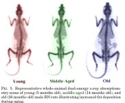
The earliest manifestations of aging are metabolic changes that result in increased fat deposition and reduced muscle mass, which lead to increased likelihood of developing "metabolic disease" (type II diabetes, hyperlipidemia, arteriosclerosis, and hypertension) (107, 108, 335). Increased fat deposition in young (5 months old), in middle-aged (14 months old), and old (26 months old) male BN rats is illustrated by dual-energy x-ray absorptiometry scans shown ibelow (336). These metabolic changes are associated with declining GH, IGF-I (see Section V), and sex steroid levels (see Section VIII) in the face of relative increases in glucocorticoid production (see SectionX), as well as insulin resistance and leptin resistance.

fig5.jpg

Anorexia is commonly associated with aging (337, 338). Normal aging is associated with a decrease in appetite and energy intake, which has been termed the anorexia of aging (339, 340). Generally, after age 70-75 yr, the reduction in energy intake exceeds energy expenditure, resulting in weight loss where loss of muscle (sarcopenia) predominates and predisposes older subjects to protein energy malnutrition (340, 341). The observed malnutrition and sarcopenia correlate with increased morbidity, mortality, and a number of hospitalizations with extended stays (342). The causes of the physiological anorexia typified during aging are unknown; they are probably multifactorial and include a reduction in feeding drive with increased activity of satiety signals.

Healthy elderly subjects apparently retain their sensitivity to the satiating effects of cholecystokinin (CCK) and have higher fasting and postprandial CCK concentrations than young adults (343, 344). Indeed, it has been reported that CCK concentrations are higher in undernourished elderly subjects compared with the healthy elderly (345). Although circulating ghrelin concentrations increase between early adulthood and middle age in humans, there is evidence that old age is associated with decreased ghrelin concentrations in rodents and in humans (311, 346). Therefore, enhanced effects of CCK and/or reduced effects of ghrelin may contribute to the development of anorexia and, in some cases, protein malnutrition during aging.

A. Aging, ghrelin, and energy balance

Ghrelin, which is mainly produced and secreted by the gastric mucosa, stimulates food intake as well as GH secretion (92, 347). It is possible that circulating ghrelin levels decline during aging because of impaired function of the gastric mucosa. Indeed, the thickness of the membrane, the length of the glands, and the number of the endocrine cells in the gastric mucosa decrease in animals between puberty and old age (348, 349). If indeed this mechanism is operative in old subjects, we must elucidate how peripheral and central components of ghrelin action are functionally interrelated.

The age-related decline of plasma ghrelin concentrations might be related to the anorexia often observed in aged subjects. However, before we can make definitive conclusions, much larger cohorts of subjects must be evaluated to support the finding that ghrelin decreases during aging.

We discussed previously that chronic treatment of elderly subjects with ghrelin mimetics restores the age-related decline in amplitude of GH pulsatility and circulating IGF-I to levels typical of young adults (25, 38, 279). These results suggest that during aging either ghrelin production declines or ghrelin resistance occurs. The orexigenic property of ghrelin coupled with its anabolic effects via the GH/IGF-I axis and its inhibition of the production of inflammatory cytokines (315) indicate that rescue of reduced GHS-R activity by treatment with exogenous ghrelin or ghrelin mimetics may prove beneficial in the anorexia of aging.

B. Ghrelin production in CNS orexigenic centers

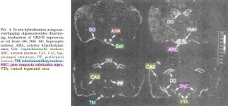
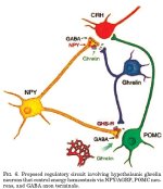
Ghrelin produced by A cells in the stomach appears to be an important peripheral orexigenic signal to the brain (350). By using a selective antibody for ghrelin and using ghrelin knockout mice as controls, the question of whether ghrelin was expressed in areas of the hypothalamus involved in regulating energy balance was addressed (294, 351). Ghrelin immunoreactive cells were identified that fill the internuclear space between the lateral arcuate hypothalamus (LAH), ventral medial hypothalamus (VMH), dorsomedial hypothalamus, paraventricular nucleus (PVN), and the ependymal layer of the third ventricle. This unique distribution does not overlap with known hypothalamic cell populations, such as those that produce NPY, AGRP, POMC, melanin-concentrating hormone, orexin, dopamine, and somatostatin 8–14. These observations suggest specific roles for locally produced ghrelin in the CNS.

Immunoelectron microscopy showed that ghrelin is located in axons where it is associated with dense-cored vesicles in presynaptic terminals (294). These axon terminals innervate the arcuate nucleus, dorsomedial hypothalamus, LAH, PVN, and ghrelin boutons and appear to make synaptic contact with cell bodies, dendrites of NPY/AGRP, POMC neurons in the arcuate nucleus, and NPY and GABA axon terminals in the arcuate nucleus and PVN. Such interactions suggest a presynaptic mode of action for ghrelin in the hypothalamus. Some ghrelin axons in the PVN innervate CRH cells, which is consistent with the increase in ACTH and glucocorticoid secretion observed following treatment with ghrelin and its mimetics. These observations delineate an anatomical basis for pre- and postsynaptic interactions between ghrelin and NPY/AGRP, POMC, and CRH circuits.

Hypothalamic localization of the GHS-R was investigated in coronal slices of rat brain using biotin-labeled ghrelin (294). Binding of biotinylated ghrelin was observed in the arcuate nucleus, LAH, and PVN was mainly associated with presynaptic boutons. Axon terminals that bound ghrelin were frequently found to contain NPY. Together, the binding data and the localization of expression of ghrelin in axons adjacent to presynaptic nerve terminals support the notion that ghrelin modulates neurotransmission.

In summary, ghrelin is produced in the hypothalamus where it is localized to a previously uncharacterized group of neurons adjacent to the third ventricle between the dorsal, ventral, paraventricular, and arcuate hypothalamic nuclei (Fig. 6) (294). These neurons send efferents onto key hypothalamic circuits, which include those producing NPY, AGRP, POMC products, and CRH. In the hypothalamus, ghrelin binds mainly to presynaptic terminals of NPY neurons. Electrophysiological recordings showed that ghrelin stimulated the activity of arcuate NPY neurons and mimicked the effect of NPY in the PVN. We propose that at these sites release of ghrelin stimulates the release of orexigenic peptides and neurotransmitters, thus representing a novel regulatory circuit controlling energy homeostasis. The involvement of NPY/AGRP neurons was confirmed by Chen and colleagues (293, 295), who showed that like Ghsr knockout mice, AGRP/NPY double knockout mice were insensitive to the orexigenic effects of ghrelin.

fig6.jpg

C. Metabolism and changes in ghrelin activity during aging

One possible explanation for altered metabolism during aging is reduced ghrelin/GHS-R signaling caused by lower production of ghrelin. Rigamonti et al. (311) found that plasma ghrelin values in old subjects (67-91 yr, n = 7) of normal weight were similar to those of young (16-36 yr, n = 7) morbidly obese, but were markedly lower than in young adults (27-39 yr, n=12) of normal weight. Therefore, because body mass index was within normal limits, an altered nutritional state was not implicated in the old subjects. The lower ghrelin levels in the old subjects were accompanied by increased insulin levels and low serum IGF-I. The former was a predicted compensatory mechanism for age-related insulin resistance, and the latter is consistent with age-dependent hyposomatotropism rather than malnutrition. Had the elderly subjects been malnourished, the low IGF-I level would have been coupled to high rather than low circulating levels of ghrelin as observed in anorexia nervosa.

Sturm et al. (352) evaluated healthy young and older women and undernourished older women. Plasma ghrelin concentrations (total active ghrelin and inactive desoctanoyl- ghrelin) were higher in undernourished older than in the well-nourished older and young subjects. Despite the fact that ghrelin stimulates appetite and food intake (92, 347), the highest circulating ghrelin concentrations were found in underweight, undernourished, older women (352). However, this does not preclude the possibility that ghrelin activity is reduced in the undernourished older subjects because of ghrelin resistance and/or increased ratio of desoctanoylated ghrelin/ghrelin. When ghrelin concentrations were compared in well-nourished young and old women, they were found to be 20% lower in older women (352). Although this difference was not statistically significant, another study evaluated a similar number of well-nourished young and old men and women and found that plasma ghrelin concentrations were significantly (~35%) lower in older subjects (311). A caveat is that although these studies suggest ghrelin production declines during adult aging, the assays used did not discriminate active ghrelin from desacyl-ghrelin.

D. Leptin, metabolism, and aging

Leptin, through its action on the hypothalamus, regulates food intake and metabolism (353–355). Mutations identified in the leptin gene of rodents and humans are associated with altered metabolism and obesity (356). Secretion of leptin is subject to ultradian pulsatile rhythmicity, although the episodic profile is not as distinct as that illustrated by pituitary hormones. However, the pulsatile pattern becomes more organized at night, where fluctuations become synchronous with those of LH and estradiol (355).

In contrast to the reproductive hormones, variations in circadian and ultradian rhythms of leptin are inversely related to ACTH and cortisol rhythms (357, 358). In vitro studies have shown that leptin regulates biosynthesis of TSH-releasing hormone, and recent studies on the synchrony of circadian/ultradian rhythms of TSH suggest that leptin also regulates TSH oscillations (359). Clearly, the compelling data in support of such a relationship do not preclude the possibility that a common pulse generator in the hypothalamus controls both leptin and TSH rhythms. The collective findings imply a permissive role for leptin in linking nutritional status and pulsatile activity of the hypothalamic-pituitary peripheral axis, they but do not prove causality.

Leptin decreases food intake and increases energy expenditure in rodents by inhibiting neurones in the hypothalamic arcuate nucleus (360). Ghrelin stimulates appetite, and its receptor (GHS-R), like the leptin receptor (Ob-Rb), is expressed in the arcuate nucleus. Ghrelin induces activation of c-fos expression in the arcuate nucleus, and 57% percent of these cells stain positive for Ob-Rb. Electrophysiology studies on hypothalamic slices show that ghrelin dose-dependently stimulates the electrical activity of these cells. Leptin is inhibitory, and ghrelin increases the electrical activity in 76% of all cells that are inhibited by leptin (360). These results show that ghrelin interacts with the leptin hypothalamic network in the arcuate nucleus and illustrate that ghrelin and leptin serve as mutual functional antagonists. Hence, ghrelin resistance can potentially be induced by increased activity of leptin and leptin-receptor in hypothalamic neurons.

E. Leptin resistance and aging

Animal models of aging have been used to investigate changes in leptin sensitivity. In rats, leptin administration selectively decreases visceral fat (VF) by approximately 60% and inhibits hepatic glucose production by approximately 80%. Surgical removal of VF improves hepatic insulin action and decreases leptin and TNF-A gene expression in sc adipose tissue (107). Therefore, the relationship between the age-related increase in VF and increased insulin resistance may involve the failure of centrally acting leptin to regulate fat distribution.

Manipulation of serum leptin levels by fasting causes hypothalamic NPY mRNA to increase in young but not in old rats (361). Leptin infusion (7 d) reduces food consumption and hypothalamic NPY concentrations by 50% in young rats; however, in old rats, food consumption is reduced by only 20% and NPY is unaffected (362). A comparison of pair-fed rats with infused with saline or leptin showed that leptin caused a 24% increase in oxygen consumption in young rats but produced no change in oxygen consumption in old rats. These results support the conclusion that aged rats are less responsive to leptin because of impaired suppression of hypothalamic NPY synthesis.

The age-related altered response to leptin has also been investigated in Zucker diabetic fatty rats, where leptin was delivered by adenovirus-mediated leptin gene transfer (361). Leptin caused markedly different responses in old (18 months old) compared with young rats (2 months old). For example, free fatty acid and triacylglycerol levels fell precipitously in the young rats but were unaffected in the old animals. Although leptin reduced food intake, body weight, and fat deposition in old rats, the effects were less pronounced than in young animals. Similarly, important metabolic markers, such as acyl coenzyme A oxidase, carnitine palmitoyl transferase-1, and peroxisome proliferator receptor A markedly increased in response to leptin in young rats but not in old rats, confirming that the beneficial metabolic effect of leptin is attenuated during aging. The mechanism of age-dependent leptin resistance is unknown. However, one possibility is that leptin receptor signaling is attenuated because of an age-dependent increase in the expression of suppressor of cytokine signaling-3 (SOCS-3) (361).

In addition to aging, leptin resistance accompanies obesity and in most cases insulin resistance. In nonobese animals, both insulin and leptin act on the hypothalamus to inhibit feeding behavior. If the anorexic action of leptin is dependent on normal insulin signaling, insulin resistance would also present as leptin resistance. To test this hypothesis, Matsumoto et al. (14) chronically administered the insulin sensitizer troglitazone (a peroxisome proliferator-activated receptor y agonist) to old BN male rats. Troglitazone reduced their high insulin, high leptin, and high body fat; furthermore, their body weight gain in response to fasting was corrected (14). Interestingly, restoration of this metabolic phenotype did not alter NPY gene expression in the arcuate nucleus. These results provide an important link between insulin and leptin resistance that apparently contributes to impairments in energy and weight regulation. Important questions must now be addressed: 1) is the mechanism independent of improved insulin sensitivity; 2) is normalization of leptin action mediated by cross talk between the insulin and leptin receptor signal transduction pathways; or 3) by improving insulin sensitivity, do asynchronous interdependent pathways essential for optimizing the biological responses to leptin become resynchronized? Clearly, additional studies are necessary to establish the mechanism of the apparent link among insulin, leptin resistance, and aging.

References:

May be found at the end of the full article: **broken link removed**
 
Last edited:
Tamoxifen reduces GH, GH pulse amplitude & IGF-1 in Normal Men

Activation of the somatotropic axis by testosterone in adult males: evidence for the role of aromatization, AJ Weissberger and KK Ho, Journal of Clinical Endocrinology & Metabolism, Vol 76, 1407-1412 1993

ABSTRACT:

To determine whether testosterone modulates the somatotropic axis in adult males, we compared 24-h GH secretion (from 20-min sampling, using Cluster analysis) and insulin-like growth factor-I (IGF-I) levels of five hypogonadal men (aged 20-32 yr) with those of six normal men (aged 21-27 yr), and examined the effects of testosterone replacement (testosterone enanthate 250 mg im monthly). To elucidate whether the action of testosterone on the somatotropic axis is direct, or requires the aromatization of testosterone to estradiol, we also examined the effects of the nonsteroidal antiestrogen, tamoxifen (20 mg/day for 3 weeks), on 24-h GH secretion and IGF-I levels in the normal men and in four of the hypogonadal men during concurrent testosterone treatment.

Compared to the normal men, the hypogonadal men had significantly reduced mean GH pulse amplitude (3.1 +/- 0.6 vs. 8.4 +/- 1.7 micrograms/L, P < 0.05), but not pulse frequency. Testosterone treatment resulted in a significant increase in 24-h mean serum GH (0.7 +/- 0.2 to 1.4 +/- 0.2 micrograms/L, P < 0.05), mean GH pulse amplitude (3.1 +/- 0.6 to 5.2 +/- 0.8 micrograms/L, P < 0.01) and serum IGF-I (0.9 +/- 0.1 to 1.1 +/- 0.1 U/mL, P < 0.05).

In the normal men, tamoxifen significantly reduced 24-h mean serum GH (1.1 +/- 0.3 to 0.5 +/- 0.1 micrograms/L, P < 0.05), mean GH pulse amplitude (8.4 +/- 1.7 to 4.7 +/- 0.4 micrograms/L, P < 0.05), and serum IGF-I (1.0 +/- 0.1 to 0.7 +/- 0.1 U/mL, P < 0.001).

In the hypogonadal men on testosterone replacement, tamoxifen lowered 24-h mean serum GH (1.3 +/- 0.2 to 0.6 +/- 0.2 micrograms/L, P < 0.01), mean GH pulse amplitude (5.5 +/- 1.0 to 2.4 +/- 0.8 micrograms/L, P < 0.01), and serum IGF-I (1.2 +/- 0.1 to 0.8 +/- 0.1 U/mL, P < 0.05).

We conclude that testosterone plays an important role in the modulation of the male somatotropic axis in adulthood, as appears to be the case in puberty, and that this effect is partly dependent on the aromatization of testosterone to estradiol.
 
Dat, I know there may not be a defenitive answer to this but what amount of either CJC and GHRP would equal out to around 4 ius of HGH a day?

Either going with straight GHRP6 can you get there or do you need to add CJC to the mix?

I was thinking it would be around 50mcg 3times a day of CJC and 100mcg of GRHP3 times per day. AM I off on that number?

I appreciate your help, you've helped out more people with this thread than any other person I've seen over the years and I would like to say thank you for that.


FYI-I'm considering getting bloodwork done to test my igf levels. My wife manages an endocrinology office so I can get it done for free. Of course my first test would be before I start this protocol, what would be the best time for the second test?
 
GHRH (Growth Hormone Releasing Hormone) + GHRP (Growth Hormone Releasing Peptide) = 10 star GH Release (**********)

GHRP (Growth Hormone Releasing Peptide aka Ghrelin-mimetic) = 3 star GH Release (***)

GHRH (Growth Hormone Releasing Hormone) = 0 or 1 star GH Release (*)


GHRPs (GHRP-6, GHRP-2, Hexarelin, Ipamorelin) are like cardiac shock paddles. You administer a GHRP and a pulse of GH is created. This is predictable and reliable across all normal people.

GHRH creates no pulse. It only adds to what ever is happening naturally. If there is a pulse occurring then GHRH increases the GH release. If no pulse is occurring when GHRH is administered then it will have little effect on GH release.​

I can not speak for Dr. Crisler but he indicated that Sermorelin (GHRH) by itself was not very effective at raising IGF-1 levels. However when he added GHRP-6 with it at saturation dose (I believe administered together twice a day), IGF-1 levels increase by 1/3.

This underscores the need for both a GHRH & a GHRP.

IF you are 100% sure you have CJC-1295 (and the odds are against it) then because it is a long lasting GHRH (half-life measured in days) it will always be available which means during natural GH waves & troughs. So it behaves differently and its effectiveness in terms of absolute GH release is higher then the other forms of GHRH.

CAVEAT - CJC-1295 raises base levels of GH not the pulses. It is possible that CJC-1295 never gives the somatotrophs sufficient time to reload stores of GH at the 100% level. Normally Somatostatin by stopping GH release activity gives the cells sufficient time to build up a big store of releasable GH. So CJC-1295 no matter how much GHRP you add may not be able to effect as strong a pulse as a GHRP + GHRH.

There is no reason NOT to combine a GHRP such as GHRP-6 with your GHRH, no matter whether the form of GHRH is Sermorelin, modified GRF(1-29) or CJC-1295. There is only BIG benefit.

On the flip side you can consistently and reliably effect GH pulsatile release with a GHRP alone. Without a GHRH the amplitude will not be synergistically higher. BUT it will be a strong pulse of GH release.

One more quick point. An iu of synthetic GH is 333mcg of compound. Thats all. A unit of GH doesn't give the same effect across all normal people and even within a person there is variability.

A far better measure is GH in plasma measured in many multiple intervals over a period of time. By sampling frequently you can determine the peak of GH in plasma and when it drops to baseline.

You can then measure IGF-1 levels to determine the effect that THAT dosing had on increasing circulating levels.

You can do the same thing with GHRH & GHRP.

The problem people have is they are stuck on absolute levels of GH in circulation as being of paramount importance. It isn't.

First it is free GH that is important. Anywhere from 10% to 90% may be bound at any given time with GH-Binding proteins or prolactin-binding proteins.

Second it is pulsation that is important for growth not absolute levels. Pulses send communicative signals to the cells. GH is simply the ligand that gives form to the wave. GH has no other value except to be a part of a communication signal.

The cells respond to this wave of GH by mediating events within the cell that are responsible for metabolism, protein synthesis, further ligand transcription & synthesis in the form of IGF-1 ...some of these signaling pathways in the cell carry messages to proliferate, differentiate or induce apoptosis . These intracellular pathways are common to many different tissue populations and respond to initiation from many different types of ligands binding to various receptors.

This behavior is optimized by pulsation ...continuous GH desensitizes these pathways (and sends certain signals that are common to females to mediate certain events such as creation of specific liver enzymes)...

So it is probable that I (and anyone who understands fully) could get more out of a small amount of GHRH + GHRP then someone who administers large amounts of GH. The validity of this statement is dependent on the use of other compounds...

Finally to answer your question directly:

I believe that if your CJC-1295 is modified GRF(1-29), coupled with GHRP-6, dosed as described you will achieve your goal of GH level (i.e. 4ius) and exceed both the quantity & quality of those growth optimizing events that THAT equivalent level of synthetic GH will be capable of mediating.


Dat, I know there may not be a defenitive answer to this but what amount of either CJC and GHRP would equal out to around 4 ius of HGH a day?

Either going with straight GHRP6 can you get there or do you need to add CJC to the mix?

I was thinking it would be around 50mcg 3times a day of CJC and 100mcg of GRHP3 times per day. AM I off on that number?

I appreciate your help, you've helped out more people with this thread than any other person I've seen over the years and I would like to say thank you for that.


FYI-I'm considering getting bloodwork done to test my igf levels. My wife manages an endocrinology office so I can get it done for free. Of course my first test would be before I start this protocol, what would be the best time for the second test?
 
Any advice would be appreciated on the downsides to doing a long acting insulin (levemir) with actual (the actual cjc-1295 peptide/ghrp-6) ?

I understand some of the pathways GH exerts itself on... need resensitizing, and insulin does that with some pathways. However hyperinsulimia is a concern of mine, but I like the logic behind doing a long acting insulin that lasts all day, for its benefits as far as stable release, less likelyhood of fat storage, avoiding those high carb meals, and the mitogenic effects.

I'm wondering how much insulin is needed over what duration of time to reduce translocation of GH receptors to the cell membrane. I understand there are a lot of variables to answering that question, but thought id put my thoughts down in words.

I was thinking maybe every other day, or 3 days on, 1 day off for a long acting insulin.

To me it sounds like there is a good synergism between a long acting cjc and a long acting insulin, but wouldn't that go down the drain if insulin caused these issues with the GH receptors and the STAT5b pathway.
 
The DHEA must significantly add to the GH if you are only taking GHRP-6 because a poster on AM first noted it by subjective feel. He was only taking GHRP-6 and added the DHEA and noticed enough of a difference to ask me about it.

**broken link removed**

Although I've seen DHEA produce such subjective benefits all on its own many times. Especially in someone who is (supposedly) HPTA-suppressed secondary to androgen supplementation. The hypogonadotropic (secondary) hypogonadism that results can dramatically lower DHEA levels. This is why I always want these gents to take some DHEA, along with PREG transdermal and low dose HCG--to backfill the hormonal pathways.
 
weltweite; said:
I'm wondering how much insulin is needed over what duration of time to reduce translocation of GH receptors to the cell membrane. I understand there are a lot of variables to answering that question, but thought id put my thoughts down in words.
...
To me it sounds like there is a good synergism between a long acting cjc and a long acting insulin, but wouldn't that go down the drain if insulin caused these issues with the GH receptors and the STAT5b pathway.

You asked a very good question.

From: Insulin Regulation of Human Hepatic Growth Hormone Receptors: Divergent Effects on Biosynthesis and Surface Translocation, Kin-Chuen Leung, Nathan Doyle, Mercedes Ballesteros, Michael J. Waters, And Ken K. Y. Ho, The Journal of Clinical Endocrinology & Metabolism 2000 Vol. 85 No. 12 4712-4720

In the study they recognized the importance of insulin and how it interacts with growth hormone, specifically highlighting that "insulin is essential for GH stimulation of IGF-I production and growth."

They then focused on the results of their study. They found that:

- Insulin up-regulated total and intracellular GH-receptors in a concentration-dependent manner.

- The abundance of GHR messenger ribonucleic acid and protein, ... respectively, markedly increased with insulin treatment.

[So the more insulin that was used the more biosynthesis or creation of GH-receptors that occurred. Now these receptors while abundant were not necessarily moved to the surface of the cell nor where they activated. Just a pool of GHRs was created.]

CAVEAT: Parts of the GH-Receptor can move to the nucleus and mediate gene expression. See the wonderful post that follows on "Growth Hormone Receptor structure, post-biogenesis behavior and degradation" :)
- It increased surface GHRs in a biphasic manner, with a peak response at 10 nmol/L, and modulated GH-induced Janus kinase-2 phosphorylation in parallel with expression of surface GHRs.

[So insulin increases the number of GH-receptors that make it to the cell surface AND increase the "intensity" of activation...but up to a point. After that point is reached insulin begins to hinder both the number of GH-receptors and "intensity" of activation"]

To quote from the study on this point:

Insulin induced a concentration-dependent increase in GHR biosynthesis, but simultaneously inhibited surface translocation. However, the net effect of reducing receptor surface availability only occurred at concentrations greater than 10 nmol/L, a concentration causing 70% inhibition of surface translocation. These data suggest that up-regulation of surface GHRs can occur with as little as 30% of intracellular receptors available for translocation to the cell surface. At concentrations above 10 nmol/L, the inhibitory effect of insulin on surface translocation overrides the compensatory effect of a 4- to 5-fold increase in receptor biosynthesis.​

[So this means that insulin increases GH-receptors by 400-500% but that as insulin rises it reduces the number of those GH-receptors that make it to the surface and are active. There is a point at which insulin begins to reduce the benefit of this GH-receptor creation. That point is 10 nmol/L of insulin. Just prior to that point insulin has inhibited substantially the translocation of GH-Receptors but the increased quantity made up for it and created an overall net benefit.]

So the problem becomes how to translate that pivot point (10 nmol/L) into a number we can use.

From: Correspondence Letter Regarding Article by von Lewinski et al, "Insulin Causes [Ca2+]i-Dependent and [Ca2+]i-Independent Positive Inotropic Effects in Failing Human Myocardium", Chih-Hsueng Hsu, MD; Cheng-I Lin, PhD; Jeng Wei, MD, Circulation. 2005;112:e367

...we find that "3 IU/L, equivalent to 20 nmol/L" ...so 10 nmol/L is equivalent to 1.5 IU/L

From Wiki Answers :D **broken link removed**

...we find that humans have 5-6 litres of blood in general.

So 5 x 1.5 = 7.5IU
So 6 x 1.5 = 9IU​

Therefore the point at which the amount of insulin in plasma becomes a negative rather then a positive is approximately 7.5 to 9 IUs.

So to arrive at a net benefit an insulin amount below that threshold point such as 5-6 ius is desirable.
 
Growth Hormone Receptor structure, post-biogenesis behavior and degradation

The following was originally created primarily for my benefit and serves as a basic summary of Growth Hormone Receptor structure, post-biogenesis behavior and degradation.

It is not intended for a wider audience and is not a Datbtrue copyrighted article. It represents the current state of knowledge in the aforementioned area (October, 2008) and was derived primarily from an unpublished paper by Stuart J. Frank & Serge Y. Fuchs on growth hormone receptor abundance and function and from Growth hormone receptor; mechanism of action, Andrew J. Brooks, Jong Wei Wooh, Kathryn A. Tunny, Michael J. Waters, The International Journal of Biochemistry & Cell Biology 40 (2008) 1984–1989

These unrefined notes are for educational use only.


GHR Structure

The growth hormone receptor (GHR) is a member of the cytokine receptor superfamily. Cytokines is a general category encompassing signaling proteins and glycoproteins (often cellular membrane proteins) that similar to hormones and neurotransmitters facilitate cellular communication. [12,13] It is a type I cytokine receptor as is the prolactin receptor, which in essence means it is connected to Janus kinase (JAK) which acts as its primary mediator of signaling events.

Physically the GHR is composed of 620 residues (a residue is an individual amino acid in a peptide chain). 350 of these residues reside inside the cell and make up what is known as the intracellular domain. 246 of these residues reside outside the cell and make up what is known as the extracellular domain. The remaining 24 residues reside in the membrane of the cell and make up what is known as the transcellular domain. [13-16]

GHRDom1.jpg

The GHR extracellular domain consists of two fibronectin type III beta sandwich domains connected by a short flexible linker.

The intracellular domain comprises Box 1 and Box 2 motifs which bind the tyrosine kinase, Janus kinase 2 (JAK2) and several tyrosine residues that act as substrates for phoshorylation by JAK2 and thus become binding sites for SH2 domain proteins.

A Kinase is a type of enzyme that transfers phosphate groups from high-energy donor molecules, such as ATP, to specific target molecules (substrates) in a process called "phosphorylation".

Since cytokine receptors such as GHR possess no catalytic kinase activity, they rely on the Janus kinase family of tyrosine kinases to phosphorylate and activate downstream proteins involved in their signal transduction pathways.

Phosphorylation of a substrate by tyrosine kinases acts as a switch to trigger binding to an SH2 domain-containing protein. STAT & PI3K proteins possess an SH2 "docking" protein and once bound begin to exert their respective effects.

The growth hormone (GH) molecule intiates this phosphorylation process by binding to one of the two GHR domains via its site of stongest attraction. For the 22kDa form of GH it would be site 1. Once bound via GH site 1 to one of the two GHR domains the GH molecule binds to the second GHR domain via the GH molecule location known as site 2.

Note that the result of the GH molecule binding with the two receptor domains does not have the effect of connecting the two receptor domains for they were already connected by a short flexible linker. Rather the binding of the GH molecule to the GHR causes a repositioning of the intracellular domains, resulting in the activation of associated tyrosine kinases and thus signal transduction. [17]

GHR2.jpg

JAK2

The GHR does not possess built-in tyrosine kinase activity and relies on associated kinases for signal transduction. The Janus kinase JAK2 provides a major component of signal transduction by GH. JAK2 associates with the GHR intracellular domain proline rich Box 1 motif via its amino terminal (JH5-7) FERM domain. JAK2 phosphorylates tyrosine residues on the associated GHR intracellular domain which provide docking sites for Src homology 2 (SH2) domain proteins, in particular STAT5a and 5b. The STATs 1, 3, 5a, and 5b are subsequently phosphorylated by JAK2, homo- or heterodimerise, translocate to the nucleus, bind to STAT responsive elements and activate transcription. [18]

GHR3.jpg

In the GHR unliganded state (unbound by the Gh molecule) it is thought that the JAK2 pseudokinase JH2 domain interacts with and autoinhibits the JH1 kinase domain resulting in an inactive JAK2. GH binding causes structural reorientation of the receptor GH domains , which results in a structural re-orientation of JAK2 and disruption of the ability of pseudokinase JH2 domains to inhibit the JH1 kinase domain, leading to JAK2 activation. There are several mechanisms for the termination of the JAK2 mediated signal which involve phosphatases, suppressors of cytokine signalling (SOCS) proteins that block signalling via their SH2 domains and receptor downregulation. [19]

In essence GH binding to the GHR triggers activation of JAK2 which causes the GHR and JAK2 tyrosine phosphorylation which induces signaling systems. The primary signaling systems are:

STATs (most notably STAT5b)
ERKs
PI3-kinase [23-25]​

GH-induced STAT5b activation requires receptor tyrosine phosphorylation and promotes gene transcription (eg., IGF-1, acid-labile subunit (ALS) of the IGF binding protein complex, SOCS proteins, hepatic P450 enzymes, and serine protease inhibitor[26–36]).

Unlike STAT5b, GH-induced ERK and PI3K activation does not require the entire GHR cytoplasmic domain, but only JAK2 coupling [23,37,38 ,39-41]. ERK activity is critical for GH-induced c-fos transcription [42], enhances GH-stimulated proliferation [43], and mediates crosstalk with EGF signaling [44-46]. GH-induced PI3K activity is implicated in antiapoptosis and/or proliferation and likely contributes to GH-induced ERK, p70 S6 kinase, and phosphodiesterase activity [42,43,47-50].

Sig1.jpg

GH sensitivity is substantially affected by the abundance of GHR available for ligand engagement at particular target cells and tissues. Surface GHR availability is regulated at several levels, including transcriptional, post-transcriptional, and post-translational.

For a thorough review of transcriptional and post-transcriptional events see references 51 & 52.

GHR regulation at the post-translational will be reviewed herein.

Continued below.
 
Last edited:
Growth Hormone Receptor structure, post-biogenesis behavior and degradation

Movement of newly synthesized GHR to the cell surface

GHR is synthesized as a non-glycosylated precursor that is transported from the endoplasmic reticulum (ER) to the Gogli apparatus. GHR dimerizes in the ER early in the process of biogenesis thus accounting for the receptor dimers detected at the cell surface even in the absence of GH engagement. [53, 60,61] In the process of transport through the Golgi, the GHR acquires carbohydrate in a characteristic fashion, in which high-mannose sugars added in the ER are ultimately removed during the transition from the early to late Golgi to yield the mature glycosylated GHR that populates the cell surface.

biogenisis.jpg

JAK2 apparently associates with GHR early in the biosynthetic pathway and not only acts as a GHR chaperone as it makes its way to the surface but fosters GHR maturation [59,62]

Studies suggest that in cells that lack JAK2 the nascent precursor GHR is a target for endoplasmic reticulum associated degradation (ERAD) and represent the first example of ERAD-associated cleavage of a cytokine receptor family member that stems from a lack of its cognate JAK. ERAD is a process whereby proteins that fail to fold properly or otherwise fail quality control mechanisms in the ER undergo retrotranslocation and proteasomal degradation in the cytosol [67–69]. JAK2, by virtue of its association with the GHR, rather than via its kinase activity, apparently "chaperones" the dimerized precursor so as to avoid quality control and proceed with efficient processing to mature GHR in the secretory pathway.

How does JAK2 exert this chaperone effect? Multiple possibilities exist, including the notion that a receptor region that might otherwise be seen as defective or unfolded to the quality control apparatus is hidden by JAK2 binding. In a similar fashion, JAK2 binding might allosterically (i.e. shape change) alter a GHR site outside of the region that interacts with JAK2 to make that site appear less defective.

In essence JAK2 association affects endoplasmic reticulum to cell surface GHR trafficking. In cells harboring GHR and JAK2 molecules that can associate, GHR moves from the ER to the Golgi, matures, and reaches the cell surface efficiently. In cells lacking JAK2 or with GHR molecules that cannot associate with JAK2 (by virtue of mutation of the receptor Box 1 region), GHR primarily undergoes endoplasmic reticulum-associated degradation (ERAD) while a small amount inefficiently matures and traffics to the cell surface (without association with JAK2).

Sig2.jpg

Proteolysis (degradation) of the GHR

Over the last decade, it has become appreciated that GHR, like some other surface receptors, is a target for regulated sequential proteolysis, the first step of which (alpha-secretase cleavage) occurs in the proximal extracellular domain stem region 8–9 residues (depending on species) outside the plasma membrane [65,80,81]. This alpha- secretase cleavage results in loss of full-length GHR, appearance of a cell-associated transmembrane domain (TMD)/intracellular domain (ICD)-containing receptor fragment (the "remnant"), and a soluble GHR extracellular domain (ECD) (called GH binding protein (GHBP)[82,83]).

GHR alpha- secretase cleavage is constitutive, but can be further induced in various cell types by a protein kinase C activator (the phorbol ester, PMA), platelet-derived growth factor (PDGF), or serum [65,80,81,84– 87]. This cleavage is catalyzed mainly by the extracellular domain of the transmembrane metalloprotease, TACE (tumor necrosis factor-alpha converting enzyme; ADAM-17) [66,88].

Importantly, inducible alpha- secretase cleavage likely regulates GH sensitivity; that is, GH-induced signaling is dampened after cells are exposed to stimuli that promote GHR alpha-secretase cleavage, but not in the presence of metalloprotease inhibitors or if noncleavable receptor mutants are expressed, suggesting that metalloproteolysis modulates GH responsiveness in part by regulating surface GHR levels [65,83,84].

Further, recent in vivo experiments indicate that administration of bacterial endotoxin leads to downregulation of hepatic GHR abundance and hepatic insensitivity to GH at least in part by inducing receptor proteolysis, suggesting that this may constitute a physiologically-relevant mechanism of regulation of GH action [89]. Notably, GH itself does not promote GHR alpha-secretase cleavage; indeed, GH inhibits subsequent GHR proteolysis, apparently by altering GHR conformation, rather than by causing signaling [66].

Recent studies have shown that the alpha-secretase-generated GHR TMD/ICD remnant is further cleaved by an enzyme activity termed gamma-secretase within the TMD, which liberates the ICD, a protein termed the "GHR stub" [87]. gamma-secretase consists of four molecules, including presenilin, which forms the aspartyl protease core and facilitates a process known as regulated intramembrane proteolysis (RIP) [90].

In essence inducible alpha-secretase cleavage generates remnant, which is converted to stub by gamma-secretase. The stub is labile and can accumulate in either the cytosol or the nucleus; proteasome inhibition prevents stub degradation.

In summary GHR undergoes sequential TACE and gamma-secretase cleavage. Surface GHR undergoes constitutive and inducible cleavage in the extracellular domain stem region by TACE in a process called "alpha-secretase" cleavage. This yields the shed GHBP and the GHR remnant. Remnant is then cleaved by gamma-secretase within the membrane to yield the GHR stub (soluble intracellular domain), which localizes to the nucleus, where it may affect gene expression.

Sig3.jpg

Surface GHR stability

Once at the cell surface, the GHR could, in principle, achieve several fates. If engaged by GH, signaling is triggered and the receptor undergoes ligand-dependent downregulation. However, in the natural milieu, GH is released from the pituitary gland in a pulsatile fashion such that GH levels are quite low in periods between pulses. Thus, it is critical to understand factors that govern GHR abundance independent of GH. It is believed that mature GHRs are cleared from the cell surface by constitutive or inducible proteolytic shedding (discussed above) and by constitutive downregulation (discussed below).

JAK2 affects the fate of the cell surface GHR and in cells lacking JAK2, the ratio of mature (cell surface):precursor GHR was substantially reduced in comparison to JAK2-replete cells [25,62]. This finding is partly explained by the chaperone effect of JAK2 during GHR biogenesis [63]. However, notable JAK2-dependent differences in the constitutive fate of mature GHRs are found as well [62]. In the context of a stable reconstitution system, the half-life (t1/2) of the receptor was estimated by anti-GHR immunoblotting after 0–4 h of treatment with cycloheximide (CHX) to inhibit new protein synthesis. The results of such a "CHX chase" assay indicated that the precursor GHR abundance dropped precipitously and to a similar degree with increasing duration of CHX treatment both in cells that did or did not express JAK2. For the mature receptor, however, there was a dramatic effect of JAK2. As measured by this assay, the GHR t1/2 increased from roughly 1 hr in cells that lack JAK2 to roughly 4 h in cells expressing JAK2 [62].

Thus, in the absence of GH, it appears that, in addition to its role in shepherding the GHR through the secretory pathway and lessening the degree to which it is targeted for ERAD, JAK2 also extends the receptor's presence at or near the cell surface, presumably by interfering with constitutively active cellular machinery that functions to internalize and downregulate the receptor.

In summary JAK2 association affects the constitutive (GH-independent) fate of surface GHR. In cells harboring GHR and JAK2 molecules that can associate, surface GHR is downregulated at a low constitutive rate and its half-life is long. In cells that lack JAK2 or have GHR and JAK2 molecules that cannot associate, GHR undergoes enhanced constitutive downregulation and exhibits a short half-life.

Sig4.jpg

GH-induced GHR downregulation

Like many surface receptors, GHR undergoes important trafficking events in response to binding of its ligand. The net effect is substantial GH-induced receptor downregulation, which serves to limit or alter the receptor's signaling capacity and perhaps thereby further emphasize the physiologic effects of pulsatile GH release from the pituitary gland. Work as early as the 1970s–1980s and since that time suggested that GH-induced GHR downregulation proceeds via clathrin coated pit-mediated endocytosis and lysosomal degradation [91,98–100].

biogenisis-2.jpg

GH ultimately causes its receptor to be degraded in lysosomes. However GH-induced receptor ubiquitination (inactivation by an attaching ubiquitin) depends on both JAK2 activity and the ability of the receptor to be tyrosine phosphorylated.

Several binding partners have been shown to associate via their SH2 domains with the tyrosine phosphorylated intracellular domain [107–111, 114]. One of them, the protein tyrosine phosphatase, SHP-2, may contribute modestly to GH-induced GHR downregulation [108]. More recently, it has been appreciated that the SOCS family protein CIS (cytokine inducible SH2 domain-containing protein), which interacts with tyrosine phosphorylated GHR [109,110] and is likely linked to Cullin5-based E3 ubiquitin ligase complex that can recruit proteins to the proteasome for degradation [112], promotes GH-induced GHR internalization and thus can desensitize GH signaling [113].

REFERENCES:

[1] - [11] - UNUSED

[12] J.F. Bazan, Structural design and molecular evolution of a cytokine receptor superfamily, Proc. Natl. Acad. Sci. U. S. A. 87 (1990) 6934–6938.

[13] D.W. Leung, S.A. Spencer, G. Cachianes, R.G. Hammonds, C. Collins,W.J. Henzel, R. Barnard, M.J. Waters, W.I. Wood, Growth hormone receptor and serum binding protein: purification, cloning and expression, Nature 330 (1987) 537–543.

[14] S.J. Frank, J.L. Messina, Growth hormone receptor, in: J.J. Oppenheim, M. Feldman (Eds.), Cytokine Reference On-Line, Academic Press, Harcourt, London, UK, 2002, pp. 1–21, Website, www.academicpress.com/cytokinereference, 24-hour free access.

[15] C. Carter Su, J. Schwartz, L.S. Smit, Molecular mechanism of growth hormone action, Annu. Rev. Physiol. 58 (1996) 187–207.

[16] S.J. Frank, J.J. O'Shea (Eds.), Recent Advances in Cytokine Signal Transduction: Lessons from Growth Hormone and other Cytokines, 1999, Greenwich, CT.

[17] Brown, R. J., Adams, J. J., Pelekanos, R. A.,Wan,Y., McKinstry,W. J., Palethorpe, K., et al. (2005). Model for growth hormone receptor activation based on subunit rotation within a receptor dimer. Nat. Struct. Mol. Biol., 12(9), 814–821.

[18] Ihle, J. N., & Gilliland, D. G. (2007). Jak2: Normal function and role in hematopoietic disorders. Curr. Opin. Genet. Dev., 17(1), 8–14.

[19] Waters, M. J., Hoang, H. N., Fairlie, D. P., Pelekanos, R. A., & Brown, R. J. (2006). New insights into growth hormone action. J. Mol. Endocrinol., 36(1), 1–7.

[20] Zhu, T., Ling, L., & Lobie, P. E. (2002). Identification of a JAK2- independent pathway regulating growth hormone (GH)-stimulated p44/42 mitogen-activated protein kinase activity. GH activation of Ral and phospholipaseDis Src-dependent. J. Biol. Chem., 277(47), 45592–45603.

[21] Rowlinson, S. W., Yoshizato, H., Brown, R. J., Behncken, S. N., Barclay, J. L., Brooks, A. J., et al. An activation-related conformational change determines signalling pathway choice for the growth hormone (GH) receptor. Manuscript submitted for publication.

[22] Fresno Vara, J. A., Caceres, M. A., Silva, A., & Martin-Perez, J. (2001). Src family kinases are required for prolactin induction of cell proliferation. Mol. Biol. Cell, 12(7), 2171–2183.

[23] S.J. Frank, J.L. Messina, Growth hormone receptor, in: J.J. Oppenheim, M. Feldman (Eds.), Cytokine Reference On-Line, Academic Press, Harcourt, London, UK, 2002, pp. 1–21, Website, www.academicpress.com/cytokinereference, 24-hour free access.

[24] C. Carter Su, J. Schwartz, L.S. Smit, Molecular mechanism of growth hormone action, Annu. Rev. Physiol. 58 (1996) 187–207.

[25] S.J. Frank, J.J. O'Shea (Eds.), Recent Advances in Cytokine Signal Transduction: Lessons from Growth Hormone and other Cytokines, 1999, Greenwich, CT.

[26] L.H. Hansen, X. Wang, J.J. Kopchick, P. Bouchelouche, J.H. Nielsen, E.D. Galsgaard, N. Billestrup, Identification of tyrosine residues in the intracellular domain of the growth hormone receptor required for transcriptional signaling and Stat5 activation, J. Biol. Chem. 271 (1996) 12669–12673.

[27] L.S. Smit, D.J. Meyer, N. Billestrup, G. Norstedt, J. Schwartz, C. Carter-Su, The role of the growth hormone (GH) receptor and JAK1 and JAK2 kinases in the activation of Stats 1, 3, and 5 by GH, Mol. Endocrinol. 10 (1996) 519–533.

[28] A. Sotiropoulos, S. Moutoussamy, F. Renaudie, M. Clauss, C. Kayser, F. Gouilleux, P.A. Kelly, J. Finidori, Differential activation of Stat3 and Stat5 by distinct regions of the growth hormone receptor, Mol. Endocrinol. 10 (1996) 998–1009.

[29] X. Wang, C.J. Darus, B.C. Xu, J.J. Kopchick, Identification of growth hormone receptor (GHR) tyrosine residues required for GHR phosphorylation and JAK2 and STAT5 activation, Mol. Endocrinol. 10 (1996) 1249–1260.

[30] W. Yi, S.O. Kim, J. Jiang, S.H. Park, A.S. Kraft, D.J. Waxman, S.J. Frank, Growth hormone receptor cytoplasmic domain differentially promotes tyrosine phosphorylation of signal transducers and activators of transcription 5b and 3 by activated JAK2 kinase, Mol. Endocrinol. 10 (1996) 1425–1443.

[31] P.L. Bergad, H.M. Shih, H.C. Towle, S.J. Schwarzenberg, S.A. Berry, Growth hormone induction of hepatic serine protease inhibitor 2.1 transcription is mediated by a Stat5-related factor binding synergistically to two alphaactivated sites, J. Biol. Chem. 270 (1995) 24903–24910.

[32] H.W. Davey, M.J. McLachlan, R.J. Wilkins, D.J. Hilton, T.E. Adams, STAT5b mediates the GH-induced expression of SOCS-2 and SOCS-3 mRNA in the liver, Mol. Cell Endocrinol. 158 (1999) 111–116.

[33] H.W. Davey, T. Xie, M.J. McLachlan, R.J. Wilkins, D.J. Waxman, D.R. Grattan, STAT5b is required for GH-induced liver IGF-I gene expression, Endocrinology 142 (2001) 3836–3841.

[34] G.B. Udy, R.P. Towers, R.G. Snell, R.J. Wilkins, S.H. Park, P.A. Ram, D.J. Waxman, H.W. Davey, Requirement of STAT5b for sexual dimorphism of body growth rates and liver gene expression, Proc. Natl. Acad. Sci. U. S. A. 94 (1997) 7239–7244.

[35] G.T. Ooi, K.R. Hurst, M.N. Poy, M.M. Rechler, Y.R. Boisclair, Binding of STAT5a and STAT5b to a single element resembling a alpha-interferon-activated sequence mediates the growth hormone induction of the mouse acid-labile subunit promoter in liver cells, Molecular. Endocrinology. 12 (1998) 675–687.

[36] J.Woelfle, J. Billiard, P. Rotwein, Acute control of insulin-like growth factor-I gene transcription by growth hormone through Stat5b, J. Biol. Chem. 278 (2003) 22696–22702

[37] A. Sotiropoulos, M. Perrot-Applanat, H. Dinerstein, A. Pallier, M.C. Postel-Vinay, J. Finidori, P.A. Kelly, Distinct cytoplasmic regions of the growth hormone receptor are required for activation of JAK2, mitogen-activated protein kinase, and transcription, Endocrinology 135 (1994) 1292–1298.

[38] J.A. Vanderkuur, X. Wang, L. Zhang, G.S. Campbell, G. Allevato, N. Billestrup, G. Norstedt, C. Carter-Su, Domains of the growth hormone receptor required for association and activation of JAK2 tyrosine kinase, J. Biol. Chem. 269 (1994) 21709–21717.

[39] C. Moller, A. Hansson, B. Enberg, P.E. Lobie, G. Norstedt, Growth hormone (GH) induction of tyrosine phosphorylation and activation of mitogen-activated protein kinases in cells transfected with rat GH receptor cDNA, J. Biol. Chem. 267 (1992) 23403–23408.

[40] L.S. Argetsinger, G.W. Hsu, M.G.J. Myers, N. Billestrup, M.F. White, C. Carter-Su, Growth hormone, interferon-alpha, and leukemia inhibitory factor promoted tyrosyl phosphorylation of insulin receptor substrate-1, J. Biol. Chem. 270 (1995) 14685–14692.

[41] L.S. Argetsinger, G. Norstedt, N. Billestrup, M.F. White, C. Carter-Su, Growth hormone, interferon-alpha, and leukemia inhibitory factor utilize insulin receptor substrate-2 in intracellular signaling, J. Biol. Chem. 271 (1996) 29415–29421

[42] C. Hodge, J. Liao, M. Stofega, K. Guan, C. Carter-Su, J. Schwartz, Growth hormone stimulates phosphorylation and activation of elk-1 and expression of c-fos, egr-1, and junB through activation of extracellular signal-regulated kinases 1 and 2, J. Biol. Chem. 273 (1998) 31327–31336.

[43] L. Liang, T. Zhou, J. Jiang, J.H. Pierce, T.A. Gustafson, S.J. Frank, Insulin receptor substrate-1 enhances growth hormone-induced proliferation, Endocrinology 140 (1999) 1972–1983.

[44] S.O. Kim, J.C. Houtman, J. Jiang, J.M. Ruppert, P.J. Bertics, S.J. Frank, Growth hormone-induced alteration in ErbB-2 phosphorylation status in 3T3-F442A fibroblasts, J. Biol. Chem. 274 (1999) 36015–36024.

[45] Y. Huang, Y. Chang, X.Wang, J. Jiang, S.J. Frank, Growth hormone alters epidermal growth factor receptor binding affinity via activation of ERKs in 3T3-F442A cells, Endocrinology 145 (2004) 3297–3306

[46] J.A. Costoya, J. Finidori, S. Moutoussamy, R. Searis, J. Devesa, V.M. Arce, Activation of growth hormone receptor delivers an antiapoptotic signal: evidence for a role of Akt in this pathway, Endocrinology 140 (1999) 5937–5943.

[47] S. Jeay, G.E. Sonenshein, P.A. Kelly, M.C. Postel-Vinay, E. Baixeras, Growth hormone exerts antiapoptotic and proliferative effects through two different pathways involving nuclear factor-kappaB and phosphatidylinositol 3-kinase, Endocrinology 142 (2001) 147–156.

[48] E. Kilgour, I. Gout, N.G. Anderson, Requirement for phosphoinositide 3-OH kinase in growth hormone signalling to the mitogen-activated protein kinase and p70s6k pathways, Biochem. J. 315 (1996) 517–522.

[49] S.J. MacKenzie, S.J. Yarwood, A.H. Peden, G.B. Bolger, R.G. Vernon, M.D. Houslay, Stimulation of p70S6 kinase via a growth hormone-controlled phosphatidylinositol 3-kinase pathway leads to the activation of a PDE4A cyclic AMP-specific phosphodiesterase in 3T3-F442A preadipocytes, Proc. Natl. Acad. Sci. U. S. A. 95 (1998) 3549–3554.

[50] L. Liang, J. Jiang, S.J. Frank, Insulin receptor substrate-1-mediated enhancement of growth hormone-induced mitogen-activated protein kinase activation, Endocrinology 141 (2000) 3328–3336.

[51] G. Schwartzbauer, R.K. Menon, Regulation of growth hormone receptor gene expression, Mol. Genet. Metab. 63 (1998) 243–253.

[52] F. Talamantes, R. Ortiz, Structure and regulation of expression of the mouse GH receptor, J. Endocrinol. 175 (2002) 55–59.

[53] J. Gent, P. van Kerkhof, M. Roza, G. Bu, G.J. Strous, Ligand-independent growth hormone receptor dimerization occurs in the endoplasmic reticulum and is required for ubiquitin system-dependent endocytosis, Proc. Natl. Acad. Sci. U. S. A. 99 (2002) 9858–9863.

[54] - [58] - UNUSED

[59] K. He, X.Wang, J. Jiang, R. Guan, K.E. Bernstein, P.P. Sayeski, S.J. Frank, Janus kinase 2 determinants for growth hormone receptor association, surface assembly, and signaling, Mol. Endocrinol. 17 (2003) 2211–2227.

[60] R.J. Brown, J.J. Adams, R.A. Pelekanos, Y.Wan,W.J. McKinstry, K. Palethorpe, R.M. Seeber, T.A. Monks, K.A. Eidne, M.W. Parker, M.J. Waters, Model for growth hormone receptor activation based on subunit rotation within a receptor dimer, Nat. Struct. Mol. Biol. 12 (2005) 814–821.

[61] M.J. van den Eijnden, L.L. Lahaye, G.J. Strous, Disulfide bonds determine growth hormone receptor folding, dimerisation and ligand binding, J. Cell. Sci. 119 (2006) 3078–3086.

[62] K. He, K. Loesch, J.W. Cowan, X. Li, L. Deng, X. Wang, J. Jiang, S.J. Frank, JAK2 enhances the stability of the mature GH receptor. Endocrinology 145 (2005) 4755–4765.

[63] K. Loesch, L. Deng, X. Wang, K. He, J. Jiang, S.J. Frank, Endoplasmic reticulumassociated degradation of growth hormone receptor in Janus kinase 2-deficient cells, Endocrinology 148 (2007) 5955–5965

[64] - UNUSED

[65] X. Wang, K. He, M. Gerhart, Y. Huang, J. Jiang, R.J. Paxton, S. Yang, C. Lu, R.K. Menon, R.A. Black, G. Baumann, S.J. Frank, Metalloprotease-mediated GH receptor proteolysis and GHBP shedding. Determination of extracellular domain stem region cleavage site, J. Biol. Chem. 277 (2002) 50510–50519.

[66] K. Loesch, L. Deng, J.W. Cowan, X.Wang, K. He, J. Jiang, R.A. Black, S.J. Frank, JAK2 influences growth hormone receptor metalloproteolysis, Endocrinology 147 (2006) 2839–2849.

[67] K. Romisch, Endoplasmic reticulum-associated degradation, Annu. Rev. Cell. Dev. Biol. 21 (2005) 435–456.

[68] A. Ahner, J.L. Brodsky, Checkpoints in ER-associated degradation: excuse me, which way to the proteasome? Trends. Cell. Biol. 14 (2004) 474–478.

[69] A. Schmitz, V. Herzog, Endoplasmic reticulum-associated degradation: exceptions to the rule, Eur. J. Cell. Biol. 83 (2004) 501–509.

[70] - [79] - UNUSED

[80] J. Alele, J. Jiang, J.F. Goldsmith, X. Yang, H.G. Maheshwari, R.A. Black, G. Baumann, S.J. Frank, Blockade of growth hormone receptor shedding by a metalloprotease inhibitor, Endocrinology 139 (1998) 1927–1935.

[81] X. Wang, K. He, M. Gerhart, J. Jiang, R.J. Paxton, R.K. Menon, R.A. Black, G. Baumann, S.J. Frank, Reduced proteolysis of rabbit growth hormone (GH) receptor substituted with mouse GH receptor cleavage site, Mol. Endocrinol. 17 (2003) 1931–1943.

[82] G. Baumann, Growth hormone binding protein 2001, J. Pediatr. Endocrinol. Metab. 14 (2001) 355–375.

[83] G. Baumann, S.J. Frank, Metalloproteinases and the modulation of GH signaling, J. Endocrinol. 174 (2002) 361–368.

[84] R. Guan, Y. Zhang, J. Jiang, C.A. Baumann, R.A. Black, G. Baumann, S.J. Frank, Phorbol ester- and growth factor-induced growth hormone (GH) receptor proteolysis and GH-binding protein shedding: relationship to GH receptor downregulation, Endocrinology 142 (2001) 1137–1147.

[85] Y. Zhang, R. Guan, J. Jiang, J.J. Kopchick, R.A. Black, G. Baumann, S.J. Frank, Growth hormone (GH)-induced dimerization inhibits phorbol ester-stimulated GH receptor proteolysis, J. Biol. Chem. 276 (2001) 24565–24573.

[86] J. Jiang, X. Wang, K. He, X. Li, C. Chen, P.P. Sayeski, M.J. Waters, S.J. Frank, A Conformationally-sensitive GHR (Growth Hormone (GH) Receptor) antibody: impact on GH signaling and GHR proteolysis. Mol. Endocrinol. 18 (2004) 2981–2996.

[87] J.W. Cowan, X. Wang, R. Guan, K. He, J. Jiang, G. Baumann, R.A. Black, M.S. Wolfe, S.J. Frank, Growth hormone receptor is a target for presenilin-dependent gamma-secretase cleavage, J. Biol. Chem. 280 (2005) 19331–19342.

[88] - [89] - UNUSED

[90] M.S. Wolfe, R. Kopan, Intramembrane proteolysis: theme and variations, Science 305 (2004) 1119–1123.

[91] G.J. Strous, P. van Kerkhof, The ubiquitin–proteasome pathway and the regulation of growth hormone receptor availability, Mol. Cell. Endocrinol. 197 (2002) 143–151

[92] - [97] - UNUSED

[98] P. Roupas, A.C. Herington, Intracellular processing of growth hormone receptors by adipocytes in primary culture, Mol. Cell. Endocrinol. 57 (1988) 93–99.

[99] M.A. Lesniak, J. Roth, Regulation of receptor concentration by homologous hormone. Effect of human growth hormone on its receptor in IM-9 lymphocytes, J. Biol. Chem. 251 (1976) 3720–3729.

[100] N. Hizuka, P. Gorden, M.A. Lesniak, E. Van Obberghen, J.L. Carpentier, L. Orci, Polypeptide hormone degradation and receptor regulation are coupled to ligand internalization. A direct biochemical and morphologic demonstration, J. Biol. Chem. 256 (1981) 4591–4597.

[101] - [106] - UNUSED

[107] S. Moutoussamy, F. Renaudie, F. Lago, P.A. Kelly, J. Finidori, Grb10 identified as a potential regulator of growth hormone (GH) signaling by cloning of GH receptor target proteins, J. Biol. Chem. 273 (1998) 15906–15912.

[108] M.R. Stofega, J. Herrington, N. Billestrup, C. Carter-Su, Mutation of the SHP-2 binding site in growth hormone (GH) receptor prolongs GH-promoted tyrosyl phosphorylation of GH receptor, JAK2, and STAT5B, Mol. Endocrinol. 14 (2000) 1338–1350.

[109] P.A. Ram, D.J. Waxman, SOCS/CIS protein inhibition of growth hormonestimulated STAT5 signaling by multiple mechanisms, J. Biol. Chem. 274 (1999) 35553–35561.

[110] L. Du, G.P. Frick, L.R. Tai, A. Yoshimura, H.M. Goodman, Interaction of the growth hormone receptor with cytokine-induced Src homology domain 2 protein in rat adipocytes, Endocrinology 144 (2003) 868–876.

[111] S.O. Kim, J. Jiang, W. Yi, G.S. Feng, S.J. Frank, Involvement of the Src homology 2- containing tyrosine phosphatase SHP-2 in growth hormone signaling, J. Biol. Chem. 273 (1998) 2344–2354.

[112] J.G. Zhang, A. Farley, S.E. Nicholson, T.A. Willson, L.M. Zugaro, R.J. Simpson, R.L. Moritz, D. Cary, R. Richardson, G. Hausmann, B.J. Kile, S.B. Kent,W.S. Alexander, D. Metcalf, D.J. Hilton, N.A. Nicola, M. Baca, The conserved SOCS box motif in suppressors of cytokine signaling binds to elongins B and C and may couple bound proteins to proteasomal degradation, Proc. Natl. Acad. Sci. U. S. A. 96 (1999) 2071–2076.

[113] T. Landsman, D.J.Waxman, Role of the cytokine-induced SH2 domain-containing protein CIS in growth hormone receptor internalization, J. Biol. Chem. 280 (2005) 37471–37480.

[114] W. Yi, S.O. Kim, J. Jiang, S.H. Park, A.S. Kraft, D.J. Waxman, S.J. Frank, Growth hormone receptor cytoplasmic domain differentially promotes tyrosine phosphorylation of signal transducers and activators of transcription 5b and 3 by activated JAK2 kinase, Mol. Endocrinol. 10 (1996) 1425–1443.​
 
Last edited:
You asked a very good question.

From: Insulin Regulation of Human Hepatic Growth Hormone Receptors: Divergent Effects on Biosynthesis and Surface Translocation, Kin-Chuen Leung, Nathan Doyle, Mercedes Ballesteros, Michael J. Waters, And Ken K. Y. Ho, The Journal of Clinical Endocrinology & Metabolism 2000 Vol. 85 No. 12 4712-4720

In the study they recognized the importance of insulin and how it interacts with growth hormone, specifically highlighting that "insulin is essential for GH stimulation of IGF-I production and growth."

They then focused on the results of their study. They found that:

- Insulin up-regulated total and intracellular GH-receptors in a concentration-dependent manner.

- The abundance of GHR messenger ribonucleic acid and protein, ... respectively, markedly increased with insulin treatment.

[So the more insulin that was used the more biosynthesis or creation of GH-receptors that occurred. Now these receptors while abundant were not necessarily moved to the surface of the cell nor where they activated. Just a pool of GHRs was created.]

CAVEAT: Parts of the GH-Receptor can move to the nucleus and mediate gene expression. See the wonderful post that follows on "Growth Hormone Receptor structure, post-biogenesis behavior and degradation" :)
- It increased surface GHRs in a biphasic manner, with a peak response at 10 nmol/L, and modulated GH-induced Janus kinase-2 phosphorylation in parallel with expression of surface GHRs.

[So insulin increases the number of GH-receptors that make it to the cell surface AND increase the "intensity" of activation...but up to a point. After that point is reached insulin begins to hinder both the number of GH-receptors and "intensity" of activation"]

To quote from the study on this point:

Insulin induced a concentration-dependent increase in GHR biosynthesis, but simultaneously inhibited surface translocation. However, the net effect of reducing receptor surface availability only occurred at concentrations greater than 10 nmol/L, a concentration causing 70% inhibition of surface translocation. These data suggest that up-regulation of surface GHRs can occur with as little as 30% of intracellular receptors available for translocation to the cell surface. At concentrations above 10 nmol/L, the inhibitory effect of insulin on surface translocation overrides the compensatory effect of a 4- to 5-fold increase in receptor biosynthesis.​

[So this means that insulin increases GH-receptors by 400-500% but that as insulin rises it reduces the number of those GH-receptors that make it to the surface and are active. There is a point at which insulin begins to reduce the benefit of this GH-receptor creation. That point is 10 nmol/L of insulin. Just prior to that point insulin has inhibited substantially the translocation of GH-Receptors but the increased quantity made up for it and created an overall net benefit.]

So the problem becomes how to translate that pivot point (10 nmol/L) into a number we can use.

From: Correspondence Letter Regarding Article by von Lewinski et al, "Insulin Causes [Ca2+]i-Dependent and [Ca2+]i-Independent Positive Inotropic Effects in Failing Human Myocardium", Chih-Hsueng Hsu, MD; Cheng-I Lin, PhD; Jeng Wei, MD, Circulation. 2005;112:e367

...we find that "3 IU/L, equivalent to 20 nmol/L" ...so 10 nmol/L is equivalent to 1.5 IU/L

From Wiki Answers :D **broken link removed**

...we find that humans have 5-6 litres of blood in general.

So 5 x 1.5 = 7.5IU
So 6 x 1.5 = 9IU​

Therefore the point at which the amount of insulin in plasma becomes a negative rather then a positive is approximately 7.5 to 9 IUs.

So to arrive at a net benefit an insulin amount below that threshold point such as 5-6 ius is desirable.


Thanks Dat, that makes sense, and it is VERY good to know, especially with the current state of how much insulin people take in general.
 

Forum statistics

Total page views
559,906,203
Threads
136,146
Messages
2,781,004
Members
160,451
Latest member
rh8767
NapsGear
HGH Power Store email banner
your-raws
Prowrist straps store banner
infinity
FLASHING-BOTTOM-BANNER-210x131
raws
Savage Labs Store email
Syntherol Site Enhancing Oil Synthol
aqpharma
YMSApril210131
hulabs
ezgif-com-resize-2-1
MA Research Chem store banner
MA Supps Store Banner
volartek
Keytech banner
musclechem
Godbullraw-bottom-banner
Injection Instructions for beginners
Knight Labs store email banner
3
ashp131
YMS-210x131-V02
Back
Top