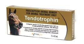
This just in! Remember this is for a horse.
What is Tendotrophin™ Injection?
Suboptimal healing, prolonged rehabilitation time
and a high incidence of recurrence make tendon
injuries difficult to treat. Tendotrophin™ Injection is
the only registered, veterinary-prescribed, equine
tendon injury treatment for use in Australian horses.
This non-surgical treatment is easily administered by
equine veterinarians. Its active constituent is Insulinlike
Growth Factor-I (IGF-I), a natural protein found in
animals and humans that is responsible for tissue
growth and repair.
The product has been developed by Primegro Limited,
after initial research conducted by the University of
Adelaide within the CRC for Tissue Growth and
Repair.
The tendinitis re-injury rate in racing horses is high, in
the range of 50-80%. Using Tendotrophin™ Injection
treatment to improve repair during the initial healing
phase, can increase the horse's chances of returning
to pre-injury performance. The two-dose course assists
injured tendons to undergo a superior repair by
ensuring the right tendon matrix synthesis occurs
early during the repair process.
How does TendotrophinTM enhance tendon healing?
Tendotrophin™ Injection increases the production of
type I collagen at the injury site, during the early
phase of repair. This significantly reduces the amount
of type III collagen produced post-injury. Type I
collagen is much thicker and stronger than type III,
which is prone to increased criss-crossing, leading to
scar tissue that weakens the tendon. Pre-injured
tendons largely contain type I collagen, therefore
Tendotrophin™ treatment aims to direct tendon
repair during the early phase of healing to produce a
tendon that has a structure similar to its original
form.
The primary benefit of Tendotrophin™ is in the early
phase of healing (first 2-3 weeks). Initial Australian
efficacy studies were conducted in the internationally
accepted collagenase-induced model of flexor
tendinitis. In two studies, treated horses had lesions
with improved sonographic healing and increased
type I collagen at 15 weeks following collagenaseinduced
injury. This demonstrates that
Tendotrophin™ treatment improves the rate of repair
and increases production of the more beneficial type I
collagen during the first three months post injury.
Independent overseas studies
Tendon healing is enhanced at the cellular level
through the direct administration of IGF-I. (Murphy
and Nixon 1997). IGF-I stimulates tenocyte
proliferation and the synthesis of type I collagen and
proteoglycans (tendon matrix). Improved mechanical
characteristics have also been shown along with antiinflammatory
effects by reducing soft tissue swelling
(Dahlgren et al 2002) post IGF-I treatment. The effects
of exogenous IGF-I can potentially lead to
significantly less tendon remodelling, which is
required to return the tendon to its mature
functional structure. Furthermore treated horses may
have an improved ability to withstand the controlled
rehabilitation program that is essential for a
successful return to performance.
Recent studies at Cornell University (Dahlgren et al
2005) have found that the levels of tendon IGF-I
become low during the early stages of tendon tissue
repair. Therefore administering IGF-I into the lesion
during the early stage of healing, seeks to boost the
low tendon tissue levels and enhance the metabolic
response of tenocytes, leading to a superior repair.
Tendon injury severity assessment
The injured tendon should be ultrasonically assessed
with the lesion size, integrity and location
determined prior to treatment and the data recorded
for comparison with future examinations. Both
transverse and longitudinal scan measurements are
essential to define the tendon lesion and tendon
integrity.
The length of the tendon lesion is determined during
the longitudinal assessment by using a 7 zone
division, every 4 cm sequentially along the tendon,
beginning from the accessary carpal bone. Some
equine clinicians prefer to record details of the
tendon lesion every 2 cm distal to the accessary carpal
bone. The fibre alignment observed in the different
zones of the lesion should be assessed.
The cross sectional area of the tendon affected
(<25%, 25-50%, >50%) is recorded for each of the
different zones during a transverse scan and these
results can be categorised providing an aid to the
horses prognosis. The position of the tendon damage
on the transverse scan is also recorded. For example;
the lesion maybe a core, a peripheral a palmar or
dorsally located lesion?
Lesions are graded on a scale of type 1 to 4 according
to the method described by Genovese et al (Table 1).
This scale assesses the severity of disruption to tendon
architecture as determined by the degree of
echogenicity.
Table 1.
Transverse ultrasound grading system used to describe
lesions in SDF tendon according to Genovese et al.
Type 1: Lesion slightly hypoechoic compared to
normal tendon.
Type 2: Lesion is half echogenic, and half anechoic
Type 3: Lesion is mostly anechoic, representing
significant fibre tearing.
Type 4: Lesion is totally anechoic, with almost total
fibre tearing and haematoma formation.
The main recording or the key figure is taken at the
point of maximal tendon damage.
A comparison of left and right limbs is recommended
to assess increase in size, fibre density and to discover
if any bilateral lesions are present.
Treatment program
It is recommended that the initial Tendotrophin™
treatment is injected as soon as practical after tendon
injury (within 21 days post injury). Initially it is
important to reduce tendon swelling and
inflammation prior to treatment. Correct support
bandaging is critical prior to, and post treatment. The
second injection (two injection course of treatment)
should be administered 7 days after the first dose.
Generally horses are box rested for the first four
weeks and hand walked. Veterinary advice specific to
each case is required for each stage of the controlled
exercise rehabilitation program.
The injections (needle 27g, 13mm attached) should be
administered into the tendon lesion following
clipping and aseptic preparation of the site. Sedation
and restraint may be necessary during the injection
procedure. Lifting the contralateral leg may help keep
the injured leg extended during administration of the
product.
The importance of an appropriate controlled
rehabilitation program is essential to the success of
tendon repair in conjunction with the course of
Tendotrophin™. Low-grade exercise improves the
longitudinal alignment of the collagen fibrils in the
repair tissue, which is essential for tendon strength.
It is important to ultrasound the tendon prior to
progressing into the next stage of the rehabilitation
program to ensure the tendon can manage increased
loading.

