
©ALL CONTENT OF THIS WEBSITE IS COPYRIGHTED AND CANNOT BE REPRODUCED WITHOUT THE ADMINISTRATORS CONSENT 2002-2024


You are using an out of date browser. It may not display this or other websites correctly.
You should upgrade or use an alternative browser.
You should upgrade or use an alternative browser.


.gif "IP Gear")


- Joined
- Nov 11, 2016
- Messages
- 210
What stage is it?
- Joined
- Oct 27, 2012
- Messages
- 9
This is exactly what I was told...
"EXAM TYPE: Lumbar spine MRI, 6/29/2017.
COMPARISON: None.
INDICATION: Intervertebral disc disorders with radiculopathy, lumbar region. Low back pain radiating of the right hip and to the knee.
TECHNIQUE: Coronal T1, sagittal T1, T2, STIR, and axial T1 and T2 images were obtained of the lumbar spine at 1.2 Tesla.
FINDINGS: There are small Schmorl's node deformities identified, particularly at L1-2 and L5-S1. Vertebral body heights are otherwise well-maintained. Alignment appears anatomic. Mild disc degeneration is present manifested primarily by T2 signal loss and slight disc space narrowing at L1-2 and L5-S1.
Sagittal images at T12-L1 and L1-2 demonstrate no evidence of a significant disc protrusion, spinal stenosis, or foraminal narrowing at these levels.
At L2-L3, no significant disc protrusion, spinal stenosis, lateral recess deformity, or foraminal narrowing is identified.
At L3-L4, no significant disc protrusion, spinal stenosis, lateral recess deformity, or foraminal narrowing is identified.
At L4-L5, no significant disc protrusion, spinal stenosis, lateral recess deformity, or foraminal narrowing is identified.
At L5-S1, disc bulging is present, asymmetric to the right. There is a superimposed, somewhat broad-based, right-sided disc protrusion/extrusion. This indents and slightly deforms the right side of the thecal sac but does not result in significant central canal stenosis. However, there is severe right lateral recess deformity and probable impingement/compression of the right S1 nerve root which may explain patient's symptoms. There is perhaps mild right-sided foraminal narrowing as well."
Conus medullaris appears intact.
IMPRESSION:
Right-sided disc protrusion/extrusion at L5-S1 associated with severe right lateral recess deformity and probable compression of the right S1 nerve root. This may explain patient's symptoms.
- Joined
- Nov 11, 2016
- Messages
- 210
A disc protrusion/extrusion isn't the worst, but you need therapy. Look into traction therapy, inversion table, swimming, etc. Disc herniations at your severity can heal on their own but need help with decompression. Schmorals nodes aren't anything bad unless they're greater than a certain size, the size wasn't noted, so I'm assuming it isn't bad, but the nodes indicate the loss of disc material, annulus fibers, and and nucleus palposus herniating into the vertebral body.
Take what people tell you on boards with a grain of salt. You need traction/decompression and core strengthening. I wouldn't jump into many reverse hyper machine without research first.
Take what people tell you on boards with a grain of salt. You need traction/decompression and core strengthening. I wouldn't jump into many reverse hyper machine without research first.
A disc protrusion/extrusion isn't the worst, but you need therapy. Look into traction therapy, inversion table, swimming, etc. Disc herniations at your severity can heal on their own but need help with decompression. Schmorals nodes aren't anything bad unless they're greater than a certain size, the size wasn't noted, so I'm assuming it isn't bad, but the nodes indicate the loss of disc material, annulus fibers, and and nucleus palposus herniating into the vertebral body.
Take what people tell you on boards with a grain of salt. You need traction/decompression and core strengthening. I wouldn't jump into many reverse hyper machine without research first.
Completely agree with this. I´ve had surgery for a herniated L4-L5 and ended up with a herniated L5-S1 plus severe degeneration and loss of disc height in the area. A Certified Chiropractor has been life changing in my case and helped me avoid surgery for the L5-S1, go and see one ASAP.
For decompression look into this, not tried it myself but it surely beats hanging upside down on an inversion table:
https://vertecorelift.wordpress.com/
Take a look here too:
https://www.foundationtraining.com/
- Joined
- Oct 27, 2012
- Messages
- 9
A disc protrusion/extrusion isn't the worst, but you need therapy. Look into traction therapy, inversion table, swimming, etc. Disc herniations at your severity can heal on their own but need help with decompression. Schmorals nodes aren't anything bad unless they're greater than a certain size, the size wasn't noted, so I'm assuming it isn't bad, but the nodes indicate the loss of disc material, annulus fibers, and and nucleus palposus herniating into the vertebral body.
Take what people tell you on boards with a grain of salt. You need traction/decompression and core strengthening. I wouldn't jump into many reverse hyper machine without research first.
Thanks for help man
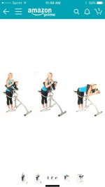
 ... I haven't been doin squats or anything to compress it more. Found this thing on amazon for $110 I'm going to give it a shot
Attachments
- Joined
- Dec 26, 2012
- Messages
- 23
I have an inversion table that works great. Good luck on ur rehab.
- Joined
- Jan 17, 2013
- Messages
- 1,417
I would avoid surgery unless absolutely necessary. In lieu of chiropractor; please consider ART. It like a chiropractor and physical therapist combined. Can really work wonders.
www.activerelease.com
BTW, are your hamstrings tight? Are you flexible?
A lot of us the problems start with flexibility. It is absolutely critical to maintain/work on flexibility training.
www.activerelease.com
BTW, are your hamstrings tight? Are you flexible?
A lot of us the problems start with flexibility. It is absolutely critical to maintain/work on flexibility training.
My discs from L2 -S1 are Fucked. I'm bone on bone from L4-S1 and the rest are bulging. It absolutely sucks and is painful. I have been staying away from pain meds for a little while now. The only thing I can't do is deadlift. I believe that working out and staying in shape keeps the surrounding muscles strong enough and protects me from injury. Keep your head up and don't over work your back.
Sent from my HTC6545LVW using Tapatalk
Sent from my HTC6545LVW using Tapatalk
- Joined
- Nov 11, 2016
- Messages
- 210
Thanks for help man
Good, avoid exercises that cause compression on the L-spine. Having axial compression, like a bar on your back, on the spine puts the most stress at L3-L5, exactly where your issues are.
That machine looks good, and looks like it simulates flexion/distraction therapy. IMO, see a chiropractor. Specifically one who has graduated from the International school or Palmer. Both are science based colleges and see if they have a flexion distraction table.
Seriously consider swimming as a form of cardio/staying active. Swimming is a full body activity, little to no spinal compression, and promotes imbition the pumping action in the intervertebral discs (IVD), nutrients in, waste out.
Remember, CORE WORK! Strengthen the anterior, posterior, lateral, and deep longitudinal chains.
I believe that working out and staying in shape keeps the surrounding muscles strong enough and protects me from injury. Keep your head up and don't over work your back.
THIS!
- Joined
- Jul 8, 2006
- Messages
- 2,409
I have compression at L3 L4 and a bulging L5 and yoga has helped alot. By loosening the hips and my t spine it has helped take pressure off the lumbar spine.
- Joined
- Jan 28, 2017
- Messages
- 253
Couldn't agree more. Everywhere you look there will be a different answers but there's a reason that acupuncture massage therapy especially deep tissue and Chiropractic Care have been around and used for so long and have hard-science backing all of them. There is probably also more confusion and disagreement on these subjects as well so I would recommend trying deep tissue massage and then going home and stretching like a bastard for a few days as well as going to a chiropractor two to three times a week for a couple of months and see if you can't get it to let up. If the chiropractor can get it to let up then you need to maintain it by stretching and possibly inversion table and massageMy discs from L2 -S1 are Fucked. I'm bone on bone from L4-S1 and the rest are bulging. It absolutely sucks and is painful. I have been staying away from pain meds for a little while now. The only thing I can't do is deadlift. I believe that working out and staying in shape keeps the surrounding muscles strong enough and protects me from injury. Keep your head up and don't over work your back.
Sent from my HTC6545LVW using Tapatalk
Sent from my SAMSUNG-SM-G870A using Professional Muscle mobile app
I have the same thing as a result of injury. It took me about 11mos to get back to 90-95%. I never skipped the gym, but found exercises that didn't directly agitate it or make it worse. I found I could rack pull with no problem...So I stuck with those. I also incorporated more back-supporting exercises when it felt funny.
My .02 is to work on core strength in a smart manner.
My .02 is to work on core strength in a smart manner.
Pilates man... get the core strong. I had to learn the hard way... heavy weight is no issue now. I've herniated l5 s1 two times. Sat in an office for years.. mixed with bad deadlift and squat form.
Sent from my SM-N920V using Professional Muscle mobile app
Sent from my SM-N920V using Professional Muscle mobile app
I was suppose to have double fusion on L3,4,5 last year, I was diagnosed with spinal stenosis, I have a bad pinched nerve, sciatica down the left leg, I could stand and was on morphine pills in real bad pain. I went to a back pain center while scheduling my fusion surgery, seeing I wouldn't be able to do this after the fusion and I heard so many horror stories of back surgery. In about 4 weeks I was much better that I cancelled my surgery. I feel the combination of chiropractics and the power decompression table was the most help. I now wear a soft lumbar support belt when I do stuff and I concentrate some of my workouts to my lower back and abs more religiously, I do hyperextensions, reverse hypers, stuff like that. I feel so much better but I do still know to be very careful now.
Similar threads
Popular tags
aas
aas testing
anabolic steroids
anabolics online
anabolid steroids
anadrol
anadrol drol tabs inj
anavar
anavar and winnie
body building
body building supplements
bodybuilder
bodybuilding
bodybuilding steroid test
clenbuterol
cycle
deca tren dosage
deca-durobolin
dianabol
dianabol and oxy
gear
hcg
hgh
motivation
muscle building
muscle mass
nandrolone
oxandrolone
pct
peptides
quality raw
raw steroid powders
steroid cycle
steroids
suspension
sust300
sustanon
test
test 400
test ace
test cyp
test cypionate
test e
test prop
testosterone
testosterone boosters
testosterone cypionate
testsuspension
tren ace
trenbolone acetate
Members online
- hemipower
- gib68sg
- samson516
- Nyoco
- Palifter
- Bloodtrail
- SOUR DIESEL
- graphics65
- gtihonov
- Twatta
- luki7788
- Dumbbell2008
- btails
- Linc0ln
- Golden
- solarsentinel1
- Musclebound94
- xyz14
- dany23x
- danieltx
- Chipper Jones78
- Drax
- Gunsmith
- nothuman
- JonnyJHutch
- warlock
- Poonami
- MOVINI711
- Yourmuscleshop_Rep1
- hhmarshall
- Pheedno
- BBSQ5
- phoenix2
- AllOkJumpmaster
- SB Labs
- powerforward
- P30LEM
- bigzzz
- Freedom69!
- ALPHALIFEFITNESS
- gojimmgo
- narta
- Hulkster74
- squatster
- Virtuous
- Blueballs
- Tank211
- chingaling
- control
- juggy38
Total: 1,311 (members: 1,303, guests: 8)
Forum statistics
- Total page views
- 559,829,875
- Threads
- 136,143
- Messages
- 2,780,852
- Members
- 160,449
- Latest member
- calebjmb


